Download

1 / 24
240 likes | 354 Vues
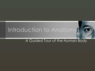
Briefing: Introduction to Anatomy of the Eye Date: 21 March 2007 Time: 1010 – 1100 . Objectives. By the end of this presentation you will be able to: Recognize major components of the eye Have a basic understanding of how the eye works

E N D
Briefing: Introduction to Anatomy of the Eye Date: 21 March 2007 Time: 1010 – 1100
Objectives • By the end of this presentation you will be able to: • Recognize major components of the eye • Have a basic understanding of how the eye works • Understand how eye disease affects the components of the eye
The Eye • The eye allows us to see and interpret the shapes, colors and dimensions of objects in the world by processing the light they reflect or emit • The eye is able to see in bright or dim light, but it cannot see objects when light is absent
The Eye When you look at any object • Light waves from that object enter the eye first through the cornea,which is the clear dome at the front of the eye • Light waves progress through the pupil, the circular opening in the center of the colorediris • Immediately behind the iris (and pupil) is the crystalline lens, and light passes through that also
The Eye • Light waves are bent (converged) first by the cornea, then even more so by the crystalline lens, to a nodalpoint which is immediately behind the lens • At the nodal point, the light waves (image) become reversed (turned backwards) and inverted (turned upside down) • Light waves continue through the vitreous humor, the clear gel that makes up about 80% of the eye’s volume, and then back to a clear focus on the retina behind the vitreous • The small, central area of the retina is the macula; it provides the best vision of any location in the retina
The Eye • The light impulses are changed into electrical signals, then sent through the optic nerve along the visual pathway to the occipital cortex, or posterior (back), of the brain • This is where the electrical signals are seen by the brain as a visual image • When light entering the eye is bright enough, the pupils will get smaller (constrict) due to pupillary light response
The Eye Extraocular Muscles
The Eye • All of the extraocular muscles, with the exception of the inferior oblique, form a “cone” within the bony orbit • The apex of the cone is in the posterior aspect (back) of the orbit, while the base of the cone is the attachment of the muscles around the midline of the eye. This conic structure is referred to as the “annulus of Zinn,” and within this cone runs the Optic nerve (cranial nerve H) • Within the optic nerve are the ophthalmic artery and the ophthalmic vein
The Eye • The superior oblique muscle is different from the others, because before it attaches to the eye, it passes through a ring-like tendon, the trochlea, which acts like a pulley in the nasal portion of the orbit • The inferior oblique muscle (not a member of the annulus of Zinn) arises from the lacrimal fossa in the nasal portion of the bony orbit and attaches to the inferior portion of the eye
The Eye • The primary muscle that moves an eye in a given direction is known as the agonist • A muscle in the same eye that moves the eye in the same direction as the agonist is known as a synergist • A muscle in the same eye that moves the eye in the opposite direction of the agonist is the antagonist
The Eye • Cardinal positions of gaze • Up/right • Up/left • Right • Left • Down/right • Down/left • In each position of gaze, one muscle of each eye is the primary mover of that eye, and is “yoked” to the primary mover of the other eye
The Eye • A “vergence” or “disconjugate” movement involves simultaneous movement of both eyes in the opposite directions • There are two principal vergence movements • Convergence – both eyes moving nasally or inward • Divergence – both eyes moving temporally or upward
The Eye Strabismus Usually when we see an object, the lines of sight are both eyes intersecting at the object, or both eyes are pointing at the object being viewed. An image of the object is focused upon the macula of each eye and the brain merges the two retinal images into one When there is an extraocular muscle imbalance, one eye is not aligned with the other eye, which results in a strabismus
The Eye Strabismus, (cont’d) With strabismus, while one eye is fixating on a particular object, the other eye is turned in another direction, either inward (cross-eyed), outward (wall-eyed), upward, or downward As a result, the person either experiences “diplopia” (double vision) or the brain learns to turn off (suppress) the image of the strabismic eye to maintain single vision The angle of deviation of the strabismus is measured in prism diopters
Diseases of the Eye Diabetic Retinopathy This is a complication of diabetes mellitus in which long-term exposure to high glucose levels in the blood has damaged retinal blood vessels. This results in new growth of abnormal blood vessels, fluid buildup in the macula (macular edema), inadequate blood supply to the retina and possibly blood and fluid leakage into the retina and the vitreous body
Diseases of the Eye • A cataract is a clouding of the eye's natural lens, which lies behind the iris and the pupil • The lens is mostly made of water and protein. The protein is arranged in a precise way that keeps the lens clear and lets light pass through it. But as we age, some of the protein may clump together and start to cloud a small area of the lens. This is a cataract, and over time, it may grow larger and cloud more of the lens, making it harder to see
Diseases of the Eye • Researchers are identifying factors that may cause cataracts such as: • People with diabetes • Users of steroids, diuretics, and major tranquilizers • Users of a lot of salt • Cigarette smoke • Air pollution • Heavy alcohol consumption
Diseases of the Eye Glaucoma • Glaucoma represents injury to the optic nerve secondary to elevated pressure inside the eye. However, there are exceptions to this definition • Some patients with sustained high intra-ocular pressure never develop any of the signs of optic nerve damage and therefore, do not truly have glaucoma. These patients are said to have ocular hypertension • Other patients may progressively lose vision and become blind, even though they never exhibit "high" eye pressures. These patients have low tension glaucoma (also called normal tension glaucoma)
Diseases of the Eye Glaucoma (cont’d) • Most patients with glaucoma do have elevated intraocular pressure. Along with the eye pressure, other parameters are evaluated in the search for glaucoma, such as peripheral vision, visual contrast sensitivity, optic nerve cupping (a hollowing out of the center of the optic nerve head in the back of the eye), and gonioscopy (visualizing the anatomy of the filtering angle of the eye, where the cornea and the iris join)
The Eye Miscellaneous tidbits about the eyes • Anterior chamber – refers to the fluid filled (aqueous humor) space between the cornea and the iris/pupil • Posterior chamber – refers to the fluid filled (aqueous humor) ring-shaped space between the iris/pupil and the lens and ciliary body • Anterior segment – refers to the intraocular portion of the eyeball and holds the anterior and posterior chambers of the eye • Posterior segment – refers to the large vitreous-filled space between the retina and the lens and where the optic nerve comes into the eye
The Eye Miscellaneous tidbits about the eyes (cont’d) • A newborn’s eyeball is about 18 millimeters in diameter, from front to back. It grows gradually to a length of approximately 24-25 millimeters (about 1”), or just smaller than a 1 1/2” ping pong ball! • CPT codes in the 60,000 series are divided by the anatomical location of either • Anterior segment • Posterior segment • Ocular adenexa
The Eye Resources: http://www.cms.hhs.gov/MLNProducts/65_ophthalmology.asp http://www.cms.hhs.gov/MLNProducts/Downloads/internet_only_manuals.pdf Internet only manuals, coding http://www.visionchannel.net/diabeticretinopathy/ Great website for information http://www.bertscope.com/Literature/Lit_Requests/info_poster.html Free eye posters and online stuff http://www.optima-hyper.com/eyetests/fitness.htm Optima’s Eye Fitness Tests