Download
1 / 33
490 likes | 1.15k Vues
Neuroendocrine cell Hyperplasia of Infancy (NEHI). Lisa R. Young, MD Director, Pediatric Rare Lung Diseases Program Cincinnati Children’s Hospital Medical Center. ChILD Foundation Conference June 25, 2011 Chicago. Overview. What are the initial symptoms of NEHI? How do we diagnose NEHI?

E N D
Neuroendocrine cell Hyperplasia of Infancy (NEHI) Lisa R. Young, MD Director, Pediatric Rare Lung Diseases Program Cincinnati Children’s Hospital Medical Center ChILD Foundation Conference June 25, 2011 Chicago
Overview • What are the initial symptoms of NEHI? • How do we diagnose NEHI? • How do we take care of children with NEHI? • What happens to children with NEHI when they get older? • Many more unknowns
NEHI is a relatively recently discovered disorder and we are still learning about it and teaching other doctors about it. • Deterding RR, Fan LL, Morton R, Hay TC, Langston C. “Persistent tachypnea of infancy (PTI) – a new entity” PediatrPulmonol, 2001. • Deterding RR, Pye C, Fan LL, et al. Persistent tachypnea of infancy is associated with neuroendocrine cell hyperplasia. PediatrPulmonol, 2005.
NEHI Symptoms • Typically presents in term infants in the first year (or months) of life • Typical symptoms in early infancy are: • Tachypnea = fast breathing • Retractions • Crackles • Hypoxia = low oxygen levels • Failure to thrive / growth difficulties Deterding et al. Persistent tachypnea of infancy is associated with neuroendocrine cell hyperplasia. Pediatr Pulmonol. 2005; 40:157-165
NEHI Symptoms: An evolving spectrum We also sometimes see kids with proven NEHI who: • were late preterm infants • have wheezing • have intermittent symptoms, more than chronic • seemed well until they had an acute illness • never need supplemental oxygen • never have growth / feeding problems
NEHI symptoms also overlap with many other causes of lung disease. • Physicians must ‘rule-out’ other more common disorders first. • But sometimes other more common problems can also complicate the picture. • Reflux • Acute infections, including viral illnesses • Occasionally patients may be misdiagnosed with NEHI.
What are the tests that enable a confident diagnosis of NEHI? • Radiology studies • Infant pulmonary function tests (infant PFTs) • Lung biopsy The term ‘NEHI Syndrome’ has been proposed for children who have chest CTs suggesting NEHI but who have not had a biopsy for confirmation.
Chest x-rays in NEHI • May be: • Normal • Hyperinflated • Look like a viral infection or bronchiolitis Deterding et al, 2005
Chest CT scans in NEHI • May be diagnostic for NEHI in many cases • Technical aspects of how the scan is done can be very important • May be atypical in at least 1/5 of children proven to have NEHI by lung biopsy (Brody et al, AJR 2010)
Chest HRCT findings in NEHI include geographic ground glass opacities, most prominent in the RML and lingula, and significant air-trapping. Importantly, no other significant abnormalities should be present. Brody et al, AJR 2010
Examples of CT scans in NEHI ‘TYPICAL’ for NEHI NOT ‘TYPICAL’ for NEHI Brody and Crotty, 2008; Brody et al, 2010; Young et al, 2010
Infant PFTs in NEHI • May aid the diagnosis of NEHI in some cases • Kerby et al (2009): • Mixed physiologic pattern in NEHI with air-trapping • Distinct from other ILD • Young et al (2010): • Severity of small airway obstruction on iPFT may correlate with extent of neuroendocrine cells seen in the biopsy • Other studies ongoing
Example of normal lung structures when viewed under the microscope
Example of normal lung from a lung biopsy when viewed under the microscope
Example of a normal bronchiole (“small air tube”) when viewed under the microscope
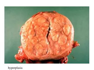
Lung biopsies look nearly normal in NEHI. Bombesin staining identifies increased numbers of neuroendocrine cells (NECs) Bronchiole with NECs Neuroendocrine body (NEB) Provided by Gail Deutsch, MD
Where are the neuroendocrine cells?In NEHI, the NEC prominence is greatest in the distal respiratory bronchioles, and distinguishes NEHI cases from other disorders associated with NEC hyperplasia. Young et al, 2010
NEHI: Variable numbers of neuroendocrine cells in different airways Deutsch et al, AJRCCM 2007
In NEHI, there is wide intra and inter-subject variability in NEC number, which does not correlate with the imaging appearance of the region biopsied. Young et al, Chest , 2010
How do we take care of children with NEHI? • There are no proven medications. • Corticosteroids (Prednisone) do not help in most cases. • Bronchodilators (breathing treatments) don’t help most long-term.
How do we take care of children with NEHI? • Supplemental oxygen when needed • Nutritional support when needed • Treat other problems as they arise, such as reflux or infections. • Avoid injuries to the lung. • Flu shot each year • Synagis to try to prevent RSV? • Avoid cigarette smoke exposure
How do we take care of children with NEHI? • Team approach • Different team members may be needed for different kids and families. • Care coordination can be very important • Psychosocial support for families • Childcare and school considerations
How do we monitor children with NEHI? • Monitoring of oxygen saturations • Monitoring of growth • Routine follow-up chest x-rays and repeat CT scans usually not helpful / not recommended. • May be needed if/when children are acutely ill or clinical course is atypical. • Infant PFTs may have a role.
What happens to children with NEHI as they get older? • Improvement over time but variable speed • May require supplemental oxygen for years • Favorable long-term outcome
What happens to children with NEHI as they get older? • At least some older children have with NEHI have abnormal PFTs and exercise intolerance. • Some exacerbate with respiratory infection. • Possible relationship to adult lung disorders is unknown. WE NEED MORE DATA! THIS IS A BIG AREA WHERE THE REGISTRY CAN HELP.
Many unanswered questions about NEHI • Why do children with NEHI often need supplemental oxygen? • What are the criteria for using oxygen in kids with NEHI?
Many unanswered questions about NEHI • Why do children with NEHI have crackles?
Many unanswered questions about NEHI • Does nutritional status and growth impact lung function in NEHI?
Many unanswered questions about NEHI • Why do some children with NEHI have ‘atypical’ chest CTs and does it matter? • Why are some children with NEHI ‘sicker’ than others?
How will we discover what causes NEHI and how to treat (or prevent) it? Genetic basis? What is the function of (or dysfunction) of neuroendocrine cells in NEHI? Are other cells involved, and what happens in lung development in children with NEHI?
Acknowledgements Rare Lung Diseases Program Connie Meeks, RN Jackie Taylor, RD Allison Whisenhunt, MISW Brenda McMillan (coordinator) Robert E. Wood, PhD, MD Bruce Trapnell, MD Pathology: Todd Boyd, DO Gail Deutsch, MD (Seattle) Radiology: Alan Brody, MD Eric Crotty, MD Mantosh Rattan, MD Pediatric Surgery: Thomas Inge, MD, PhD Infant PFTs: Jim Acton, MD (Missouri) Karen McDowell, MD Beth Koch, RRT Children’s Interstitial Lung Disease Research Network