Download
1 / 53
540 likes | 630 Vues
Learn the essential sequences for optimal liver MRI imaging, including T1-w, T2-w, diffusion, and injection sequences. Understand the principles and indications for each sequence to evaluate liver focal lesions and biliary system abnormalities effectively. Enhance your MRI technique comprehension to adapt protocols for individual patients and diseases.

E N D
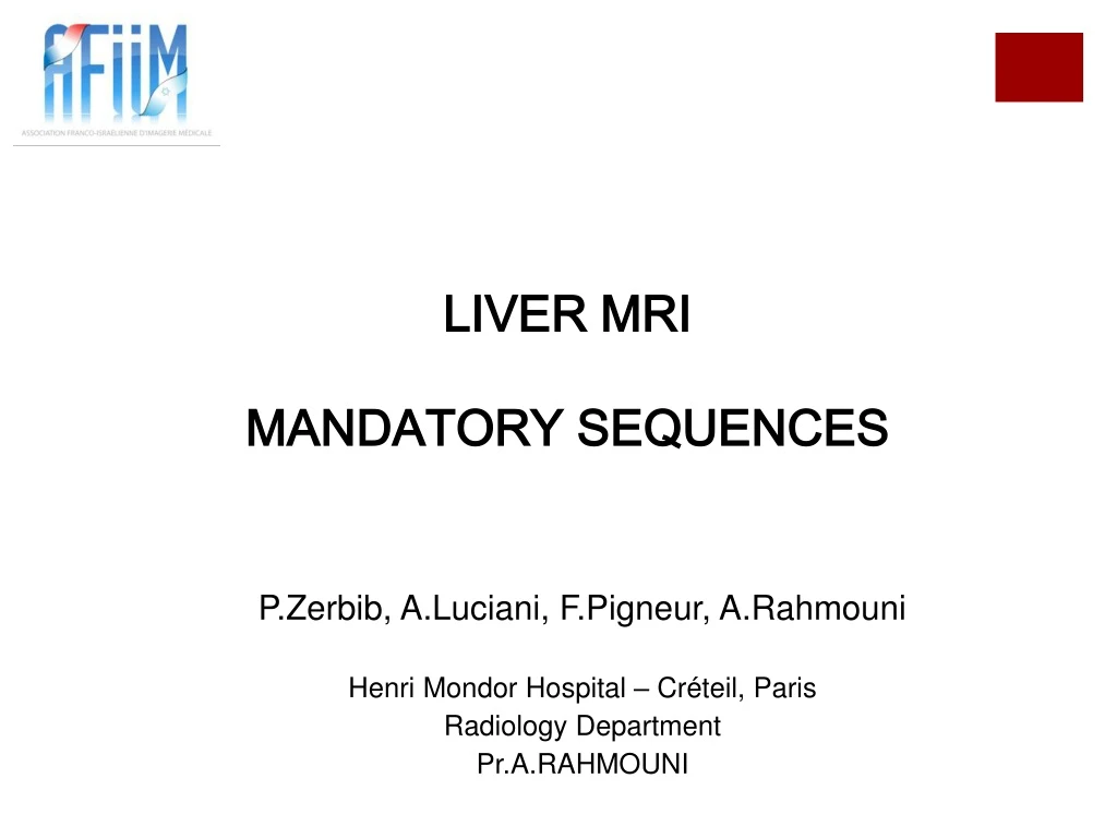
LIVER MRI MANDATORY SEQUENCES P.Zerbib, A.Luciani, F.Pigneur, A.Rahmouni Henri Mondor Hospital – Créteil, Paris Radiology Department Pr.A.RAHMOUNI
Objectives • Define a standardized protocol for the evaluation of : • Liver focal lesions • The Biliary system
Mandatory sequences to perform a good liver MR T1-w T2-w Diffusion Injection
Mandatory sequences to perform a good liver MR T1-w ? T2-w ? Diffusion ? Injection ?
PRINCIPLE 63.5Mhz E G E Fat Water G TE=4.8 ms (1.5T) TE=2.4 ms (1.5T) 220 HZ (1.5T) Fat poor substance No SI decrease in out of phase sequence TE=2.4 ms (Out of phase) TE=4.8 ms (In of phase) Fat rich substance (steatosis) SI decrease in out of phase sequence
PRINCIPLE T2-w sequences allow the detection of liver lesions (water rich substances) Time of echo= 70-100 ms
PRINCIPLE SE FastSE TR 100ms TR TR 100ms TR 100ms TR TR 100ms TR 100ms 20ms K space K space 40ms 60ms 80ms 100ms
PRINCIPLE This sequence is performed with spectral fat suppression
PRINCIPLE First echo < 70ms : DETECTION Second echo >100ms: CHARACTERIZATION long First echo Signal
PRINCIPLE First echo < 70ms : DETECTION Second echo >100ms: CHARACTERIZATION long First echo Second echo Signal
PRINCIPLE First echo < 70ms : DETECTION Second echo >100ms: CHARACTERIZATION long First echo Second echo Signal Liver Hemangioma Signal Liver-lesion contrast increment Liquid/angiomatous lesions T2-w Hepatic T2 Time of echo First echo Second echo
The purpose of diffusion sequences is to: 1. Detect small lesions hard to see in the others sequences 2. Characterize diffusion zones of restricted diffusion (eg: metastasis) from those with non restricted diffusion (eg: kystic lesions) PRINCIPLE
Principles of diffusion Restricted Diffusion Unrestricted diffusion
In the zones of restricted diffusion, signal intensity increase when diffusion gradient intensity (b) increase PRINCIPLE 180° Diffusion gradients (b = s/mm²) EPI-SE SIGNAL INTENSITY Free range of motion protons Restricted range of motion protons
b10 b20 bo b100 b30 b50 b800 b200 b400 b800
SIGNAL INTENSITY RESTRICTED DIFFUSION ELEVATED DIFFUSION b (s/mm²) ADC = (1/b1-b0) ln (S[b1]/S[b0])
ADC high signal intensity = no diffusion restriction = non suspect lesion ADC low signal intensity = diffusion restriction = suspect lesion
b50 ADC b800 Signal Analysis b50 ADC b800
b50 b400 b800 ADC b50 ADC b800 Benign lesions Suspect lesions «Shine through» T2 effect Air Présence de fer b50 b800
FAST GRADIENT ECHO T1 3D SEQUENCES WITH FAT SUPPRESSION BEFORE AND AFTER CONTRAST MEDIUM ADMINISTRATION
PRINCIPLE • GE 3D T1 : • Rapid acquisition • K space partial filling • T1-weighted images • Gadolinium injection Arterial Before Gd Portal Venous Focal Nodular Hyperplasia (FNH)
2. Biliary system abnormalities detection and/or characterization
Two sequences to study the biliary system: 1. Half-Fourier acquisition single-shot turbo spin-echo T2-weighted (HASTE) Liver and pancreatic morphologic study and biliary tract exploration 2. 2D or 3D RARE sequence (long TE) Biliary system global visualisation
PATIENT PREPARATION Patient fasting (to avoid gastric and duodenal artifacts) Fasting No fasting
HALF-FOURIER ACQUISITION SINGLE-SHOT TURBO SPIN-ECHO (HASTE)
Half Fourier acquisition (Turbo Spin Echo T2 Single shot) Half symmetric reconstruction PRINCIPLE Axial and coronal acquisitions K space symmetry
RARE SEQUENCE (BILI)(RAPID ACQUISITION RELAXED ENHANCED) PRINCIPLE
Turbo spin echo sequence with very high TE (>1000ms) Signal intensity decrease with TE High slice thickness (30 à 50 mm) Signal intensity Signal TE TE (ms)
Conclusion: • Technique comprehensionismandatory to adapt MRI protocole for each patient and eachdisease • Gradients qualityimprovements and new technologies permit to improve the diagnostic degree and to reduce the acquisition time
,מנתח בבית חולים מונדור בפריז, אני מודה אילון להט שעזר לי להכין אתהפרזנטציה הזו ב עברית
Explain the exam and reassure the patient Taking some time to explain the essentials of the upcoming MR examination process allows to save many minutes during the examination and decreases the rate of failures Motivate the patient : “The pictures could be fuzzy”, “I will talk with You during the exam”, … Explain that some sequences need breath-hold, and on the opposite that some sequences will require a regular breathing PATIENT PREPARATION
1. 2D GE T1-w sequence in and out of phase (In-Phase – Out-of-Phase) To detect a signal intensity decrease in the out-of-phase acquisition due to liver fat content 2. Fast SE T2-w sequence with spectal fat suppression / HASTE To increase the signal intensity difference between liver and lesion To explore the intra-hepatic biliary system and the principal biliary tract 3. Diffusion sequence To detect and characterize hepatic lesions 4. Fast 3D GE sequence with spectral fat suppression before and after contrast medium administration (With parallell reconstruction techniques such as Caipirinha) To study liver parenchymal enhancement and liver vascularization
Why not a gradient echo T2 acquisition? Gradient Echo Spin Echo Signal to noise ratio decrease in Gradient Echo because of liveriron content (susceptibilityartifact)
Why not a Spin Echo T1 acquisition? 180° Gp Gl Signal intensity • In and Out of phase sequence use short TE (2 - 4 ms) • Spin Echo sequence is not used because of 180° 90° 90° GE SE impulsions Gsc Gs Gp Gl Signal intensity TE TE
Toronto Consensus conference (2008) recommend: • b values between 100 and 750 s/mm² (to remove low b signal due to microperfusion) • at least 3 b values utilization, included b0, b ≥100 s/mm² and b ≥ 500 s/ mm²
Low b values evaluate the microcirculation b10 b20 bo b100 b30 b40 b800 b200 b400
Toronto Consensus conference (2008) recommend: • b values between 100 and 750 s/mm² (to remove low b signal due to microperfusion) • at least 3 b values utilization, included b0, b ≥100 s/mm² and b ≥ 500 s/ mm²
Toronto Consensus conference (2008) recommend: • b values between 100 and 750 s/mm² (to remove low b signal due to microperfusion) • at least 3 b values utilization, included b0, b ≥100 s/mm² and b ≥ 500 s/ mm²
b10 b20 bo D* D b100 b30 b800 b200 b400
Toronto Consensus conference (2008) recommend: • b values between 100 and 750 s/mm² (to remove low b signal due to microperfusion) • at least 3 b values utilization, included b0, b ≥100 s/mm² and b ≥ 500 s/ mm²
bo b10 b20 b30 b50 b100 b200 b800 b400
GE T1-w sequence with fat suppression Fat suppressed GE T1-w realized after dynamic acquisitions: Use fat suppression to increase focal enhancement axial coronal • To study parenchymal liver enhancement after Gadolinium injection • To show late focal enhancement
bo b10 b20 b30 b50 b100 b200 b800 b400