Download

1 / 81

E N D
Image Processing MRI(Magnetic Resonance Imaging) Presented by :Sara Waleed Mohamed Khames Communication &Electrons Department 1
MRI VS CT MRI: • • • CT: • • • • Imaging organs, soft tissue an internal structures (see spine scan image to the right) Showing tissue difference between normal and abnormal Imaging without radiation Imaging bone, soft tissue and blood vessels at the same time Evaluating lung and chest issues (see lung scan image to the right) Detecting cancers. Imaging patients with metal (no magnet) 4
MRI VS CT MRI: Take real section in all directions X,Y,Z, real image . CT: Take perpendicular imag and reconstruct the image in other directions. 5
MRI is suited for soft tissues evaluation: • Tumors(e.g. brain tumor). • Spinal cord disorder. • Blood vessels injuries & Aneurysms. • Strokes • Joint & Tendon injuries 6
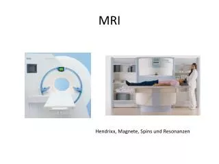
MRI Machine The main parts of the machine are: • RF Coils • Gradient Coils • Magnet 7
MRI Principle • • Two basic principles of NMR: • 1.Atoms with an odd number of protons or neutrons have spin . • 2.A moving electric charge, be it positive or negative, produces a magnetic field. MRI is based on the principle of nuclear magnetic resonance (NMR) 8
Proton • • Protons possess a positive charge. Like the earth they are constantly turning around an axis and have their own magnetic field. Fig: 1 9
Protons • Normally protons are aligned in a random fashion (Protons randomly oriented cancelled each other( Fig 2)). This, however, changes when they are exposed to a strong external magnetic field (Fig 3). • Then they are aligned in only two ways, either parallel or antiparallel to the external magnetic field. (Fig 3). 10
net magnetic moment = zero (Fig 2) (Fig 3) 11
Effect of Strong magnetic field on proton • spins align parallel (lower energy) or anti-parallel(higher energy) to B. • Number of protons parallel to B greater than the anti-parallel ones. • Opposite spins cancel the effect of each other. • Net magnetization M that is parallel to main magnetic field B. 12
Precession • Protons perform a wobbling type of motion in a strong magnetic field ,called precession. 14
Precession Frequency • • • To calculate the precession frequency. Larmor equation: ? = ??0 ?0= is the strength of the external magnetic field, which is given in Tesla (T) • ? : is the angular frequency, and ‘?’is the so- called gyromagnetic ratio. 15
Resonance & Excitation • When a nucleus is exposed to an external electromagnetic energy that has a frequency similar to its own processional frequency , the nucleus again energy from the external force. • Energy at the processional frequency of hydrogen corresponds to the radio frequency band of electromagnetic spectrum . • The application of RF pulse that cases resonance is termed excitation 16
Resonance & Excitation This absorption of energy causes an increase in number of spin down hydrogen nuclei populations as some of Spain up nuclei gain energy. As the field strength increases , the energy difference between the two population also increases so that more energy (higher frequency) are required to produce resonance. 17
The result of resonance 1-NMV moves out of alignment away from B0: The angle to which the NMV movies out of alignment is called the flip angle . The magnitude of the flip angle depends upon the amplitude and duration of the Rf pulse . B0 is now term the longitudinal axis/ plane. The plane at 90° to B0 is termed the transverse plane . e.g flip angle is 90° :nuclei is given enough energy so that the longitudinal NMV is completely transferred into transverse NMV(rotates in the transverse plane at larmor frequency) 18
The result of resonance 2-The magnetic moments of hydrogen nuclei move into phase with each: Magnetic moments become in the same place on the processional path around path around B0 at any given time. 19
MR Signal As the result of resonance , the NMV is precessing in phase in transverse plane. Faraday`s law of induction state that is a receiver coil is placed in the area of a moving magnetic field a voltage is induced in this receiver coil. A receiving coil is put in transverse plane and a voltage is produced in the coil when in the in- phase NMV precesses in the transverse plane . The coil voltage =MR signal . the frequency of the signal is the same as the larmor frequency. The magnitude of the signal depends on the amount of magnetization present in the trasverse plane. MR signal is alternating. 20
When RF switched off ,two processes occur at the same time but independently: 1)Recovery (Relaxation): • The amount of magnetization in the longitudinal plane gradually increases (the NMV is again influenced by B0) • This occur due to relaxation (i.e losses of absorb energy) 2) Decay • • the amount of magnetization in the transverse plane gradually decrease. Occur due to dephasing . 21
How can I receive signal? As the magnitude of transverse magnetization decreases, so does the magnitude of the voltage induced in receiver coil . The reduced signal is called the free induction decay (FID) Signal. 22
Principles of Magnetic Resonance Imaging There are three steps involved in identifying where in a 3D location a signal is arising from: - 1 Slice selected along z-axis - 2 Segment of slice along x-axis selected by frequency encoding - 3 Part of segment along y-axis selected by phase encoding Slice selection The first part of localizing the signal is to localize it the location of the axial slice within the object being imaged. This is known as "slice selection". The way this is done is by using the RF pulse to select which slice to activate i.e. which slice will have the magnetic vector of its nuclei flipped to the transverse plane in order to return a signal. 1. Apply gradient A magnetic field gradient is applied in the Z-axis superimposed on the background magnetic field. Going back to the Larmor equation the frequency of precession depends on the magnetic field. This means that nuclei will have different frequencies throughout the z-axis. 23
2. Select slice An RF pulse is applied to flip the magnetisation of the nuclei into the transverse plane and, therefore, give a signal. Remember, to flip the precession of the nuclei the RF pulse frequency should be the same as the Larmor frequency of the nuclei. As the Larmor frequency of nuclei is different along the z-axis we can select a slice to activate by altering the frequency of the RF pulse. 24
3. Reset As the frequencies are different along the gradient, the nuclei begin to precess out of phase. Before selecting the next slice we need to reset the nuclei. This is done by temporarily reversing the gradient to reverse the precessional frequencies. The nuclei then rephase. Summary A magnetic field gradient is applied in the z-axis The Larmor frequencies of the nuclei vary along the z-axis An RF pulse with a frequency matching the Larmor frequency of the nuclei we want to select is applied In this way, a slice along the z-axis is selected (correlates with an axial slice of the patient) The phases of the nuclei are reset by reversing the gradients 25
Spatial encoding Unlike in CT and plain films in which localization of the signal is simple (an x-ray beam travels through the material and where it hits the receptor is the physical location of what it has passed through) MRI is much more complicated. With MRI the signal is localized in the 3D space by manipulating the magnetic properties of the nuclei in a predictable way. The signals are then returned with a particular frequency and phase and these are slotted into their respective locations. The brightness of the pixel is the amplitude of the signal returned. The key concept of spatial encoding is the use of gradients. 26
Phase encoding 1. Apply phase-encoding gradient If we take a single column through the slice (i.e. a single frequency in the read-out direction) and then divide this into sections in the y-axis, each segment will have the same frequency and phase. We switch on the phase-encoding gradient along the y-axis before the readout gradient. Some sections will precess with a quicker frequency and some with a slower frequency. When we switch off the gradient all the segments return to the same frequency but they are now all out of phase with a phase-shift that depends on the position along the column. 2. Repeat cycle With every cycle the amplitude of the phase encoding gradient is changed so that the phase of the MR signal at a certain y-axis position changes each time. If we look at a single point in time along the cycle and look at the amplitude of the wave at that point, it varies with each cycle. If we then plot this amplitude with each cycle we get another wave. Each point along the segment will have a phase encoding curve with a different frequency. Those at the furthest ends of the gradient will have a greater change in their phases and, therefore, a higher frequency phase encoding curve. The point at the middle of the gradient, the isopoint, will never change its frequency or, therefore, its phase. A Fourier transform is again applied to extract these frequencies and place them in the correct place along the y-axis. The signal strength (brightness) is given by the maximum amplitude of the phase-shift curve as this corresponds to the maximum amplitude of the original signal. 28
Frequency encoding Read-out gradient We overcome this by applying another magnetic gradient in the x-axis. This changes the Larmor frequencies of the nuclei in a gradient along the x-axis. Each segment will now return a signal of a different frequency depending on its location in the x-axis. As they are of different frequencies, they will eventually become of different phases. Adding the signals together gives a large signal at the start, when they are still all in phase, but this signal drops off as the phases diverge. This gradient is called the "read out" or "frequency encoding" gradient. 29
Decoding the signal Now we have one MR signal formed of many MR signals of different frequencies. Luckily, we can easily extract out each frequency and its amplitude mathematically using the Fourier Transformation (FT). We can now map each signal to its location in the x-axis of the slice by its frequency and its amplitude (brightness). 30
K-space As we acquire our images, what we are acquiring is many, many waves of varying spatial frequency and directions. As we overlay these on top of one another we form the physical image. These wave signals are stored in stored in K-Space (aka Fourier Space) along a coordinate system. Each column of k-space contains the data obtained during one frequency encoding step. Each row is filled in by repeating the phase-encoding steps. The important things to note are: •Any particular point on K-space contributes to the whole image •Any image pixel is derived from the whole of K-space •K-space is symmetrical Within K-space the high-frequency signals are within the periphery and the low-frequency signals are within the center. The x and y-axes determine the orientation of the signal wavelengths. 33
A low-pass filter that only includes the center of K-space produces an image that is very smooth but lacks the edges and details. 35
A high-pass filter that only includes the periphery of K-space produces an image that has very good detail and edges but no low-contrast features 36
Gradient 37
Principle of Good image T1 Recovery: • The recover of longitudinal magnetization. • Caused by the nuclei giving up their energy to the surrounding environment or lattice • Often termed spin lattice relaxation. • The rate of recovery of longitudinal magnetization is an exponential process . • Recovery time constant called T1=time it takes 63% of the longitudinal magnetization to recover. 38
T2 Decay: The decay of transverse magnetization Cased by nuclei exchanging energy with neighboring nuclei >>>dephasing Often term spin spin relaxation. T2 relaxation time of tissue(time constant of decay ):the time it takes 63% of transverse magnetization to be lost N.B.A signal is only induced in the receiver coil if there is magnetization in the transverse plane , that is in phase (the signal in the coil here will decays). • • • • • 39
Pulse Timing parameter Pulse sequence consists of several component: The repetition time (TR) Is the time from application of one RF pulse to the application of the next RF pulse. Determines the amount of relaxation that is allowed to occur between the end of one RF pulse and the next of application . Therefore the TR determines the amount of T1 relaxation that has occurred. 40
The echo time (TE) Is the time from application of RF pulse to the peak of the signal induced in the coil . The TE determine how much decay of transverse magnetization is allowed to occur before the signal is read . Therefore the ,the TE controls the amount of T2 relaxation that has occurred . 42
Fat & Water hydrogen MRI characteristic Fat & water difference 1- larmor frequency; Water is hydrogen linked to oxygen which tends to steal the electrons away from around the hydrogen nucleus H is more available to the effects of the main magnetic field. Fat Is hydrogen linked to carbon ,which doesn't take electrons from around the hydrogen nucleus electron cloud protect the nucleus from the effect of the main field . Results: larmor frequency of hydrogen in water is higher than hydrogen in fat. 43
2) T1 Recovery - Fat Slow molecular tumbling in fat T1 process is relatively repaid magnetic moments of fat nuclei regain their longitudinal magnetization quickly . Results: T1 time of fat is short (100-150ms) • Results: • 44
2) T1 Recovery - water In water molecular mobility is high resulting in less efficient T1 recovery the magnetic moment take longer to relax and regain their longitudinal magnetization. Results: T1 time in water is long (1500-2000ms) • Results: • 45
3) T2 Decay Fat • Energy exchange between nuclei is more efficient in the hydrogen in fat more repaid deshaping Result: T2 time of fat is short (80ms) Water Energy exchange in water is less efficient than in fat Slow deshaping • Result: T2 time of water is long (200ms) 46
Causes of Causes of constrst constrst between tissues in MRI images between tissues in MRI images T1 contrast • T1 time of fat is shorter than water the fat vector realigns with B0 faster than water the longitudinal component of magnetization of fat is larger than water at a given time. 47
After a certain TR, the next 90 RF excitation pulse is applied the longitudinal component of magnetization of both fat & water into the transverse plane are flipped. 48
Saturation After the 2nd90° RF NMV is pushed beyond 90° (Short TR) It is said to be partially saturated T1 WT image 49
T1 weighted image • As there is longitudinal magnetization in fat before the RF pulse there is more transverse magnetization in fat after RF pulse Fat therefore has a high signal and water has low signal ion a T1 contrast image. 50