
SlideServe
Menu
Préférences
© SlideServe · v2026

MRI Findings

merrill avatar
6 diapositives · 227 Views · Jul 19, 2014

Review of severe hyperintensity in the lateral branch of the SDF tendon, PIP joint effusion, and soft tissue thickening, post-shockwave therapy, progressing well without lameness. Analysis of tendon injury and joint effusion interaction.
MRI Findings - slide 1
01 / 6MRI Findings
1 / 17

Télécharger la présentation

MRI Findings
Une image / un lien ci-dessous est fourni (tel quel) pour télécharger la présentation

Politique de téléchargement : le contenu du site vous est fourni TEL QUEL pour votre information et votre usage personnel ; il ne peut être vendu, concédé sous licence ni partagé sur d’autres sites sans l’accord de son auteur.Le contenu vous est fourni TEL QUEL, pour votre information et votre usage personnel uniquement. Téléchargez la présentation en cliquant sur ce lien. Lors du téléchargement, si vous ne parvenez pas à télécharger une présentation, l’éditeur a peut-être supprimé le fichier de son serveur.Pendant le téléchargement, si vous ne parvenez pas à obtenir une présentation, le fichier a peut-être été supprimé par l’éditeur.

F I N

Plus de contenu associé

Transcription de la présentation
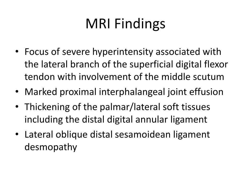
  1. MRI Findings • Focus of severe hyperintensity associated with the lateral branch of the superficial digital flexor tendon with involvement of the middle scutum • Marked proximal interphalangeal joint effusion • Thickening of the palmar/lateral soft tissues including the distal digital annular ligament • Lateral oblique distal sesamoidean ligament desmopathy

  2. Follow-Up • This imaging was performed in early December 2012. Since that time the horse was rested further, and recently the tendon lesion was treated using shockwave therapy. • Following therapy the swelling decreased and the patient is at the trotting stage of rehabilitation with no signs of lameness

  3. Commentary • This case was chosen because of the severe injury to the distal superficial digital flexor tendon and scutum in combination with pastern joint effusion • It is suspected that the peripheral, dorsal nature of the tendon injury may have resulted in hemorrhage into the pastern joint • Due to the severity of the swelling in the area joint sampling was not performed following imaging in this case

  4. Commentary • It was also interesting to see a case of severe lameness following PDN block and distal interphalangeal joint injection • No unusual events or limb retraction took place during this block • It is believed that a mild injury to the tendon was pre-existing and was exacerbated following turn-out after the exam (while the area was still under the influence of local anesthesia)