Download
1 / 21
210 likes | 336 Vues
Explore the Tissue Level of Organization, including the categories of tissues, functions, and examples of epithelial tissues such as simple squamous, cuboidal, and columnar. Learn about basement membranes, surface areas, and cellular arrangements in various epithelial tissues.

E N D
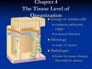
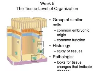
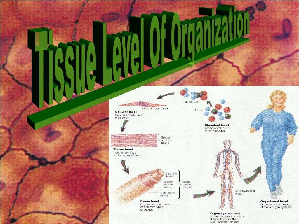
The Tissue Level of Organization • Group of similar cells • common embryonic origin • common function • bound together by intercellular substance • Histology • study of tissues
The Origin of Tissues Morula Blastula Gastrula
4 Basic Tissues Categories • Epithelial • Connective • Muscle • Nervous
Epithelial Tissue -- General Features • Cover surfaces, line cavities and form glands • FUNCTION – To create tissue that line, cover, protect, secrete. Tend to act as a barrier or covering. • Attached to underlying connective tissue by a basement membrane. • Cells are tightly packed together, to form tight network. • Avascular---without blood vessels • nutrients diffuse in from blood vessels in underlying connective tissue • What does this mean for especially thick epithelia like skin? • Rapid cell division; responsive to environmental stresses. • Named according to the SHAPE and ARRANGEMENT of cells.
SimpleSquamous Epithelium • Single layer (= SIMPLE) of flat cells (Squamous). • lines blood vessels, form capillary walls, form alveoli. • VERY THIN LAYER--. What is the primary function of this tissue?
Section through lungs Why would simple squamous be a good design for alveoli ? Surface view of lining of peritoneal (abdominal) cavity Examples of Simple Squamous Epithelium
SimpleCuboidal Epithelium • Single layer of cube-shaped cells viewed from the side. • Cuboidal cells usually are found lining/forming gland ducts.
Example of Simple Cuboidal Epithelium • Sectional view of kidney tubules.
Nonciliated SimpleColumnar • Unicellular glands =goblet cells secrete mucus • lubricate GI, respiratory, reproductive and urinary systems. • Microvilli = fingerlike cytoplasmic projections on the cell membrane’s top surface. Microvilli are designed to increase surface area to increase absorption.
Example Nonciliated Simple Columnar Epithelium • Section from small intestine The term “brush border” is often used to describe this tissue. Why is having increased surface area along surface of small intestine a good design?
CiliatedSimpleColumnar Epithelium • Single layer of rectangular cells with cilia, cilia are locomotive structures that consist of a 9 +2 arrangement of microtubules that are covered with cell membrane. • Mucus from goblet cells moved along by cilia. • Why would we want the bronchi and bronchioli lined with this type of tissue?
Example Ciliated Simple Columnar Epithelium • Section of fallopian tube. • Why would we want this structure lined with cilliated columnar cells?
Stratified Squamous Epithelium • Several cell layers thick • Surface cells flat • Keratinized = surface cells dead and filled with Keratin protein • Examples include the skin (epidermis) and mucous membranes (non-keratinized) What do you think the the primary function of this type of epithelium is all about?
Example of Stratified Squamous • Section of vagina
Stratified Cuboidal Epithelium • Multilayered • Surface cells cuboidal • rare (only found in sweat gland ducts & male urethra)
Stratified Columnar Epithelium • Multilayered • Surface cells columnar • Rare (very large ducts & part of male urethra)
Multilayered Surface cells varying in shape from round to flat if stretched Lines hollow organs of the urinary tract that expand from within. Perfect example is the urinary bladder Transitional Epithelium
Single cell layer All cells attach to basement membrane but not all reach free surface Nuclei at varying depths Pseudostratified Columnar PSEUDO = FALSE Non-Ciliated version found in part of male Urethra and in Vas Deferens Ciliated version found in Trachea