Exploring Diffusion and Microscope Techniques in Cellular Biology Experiments
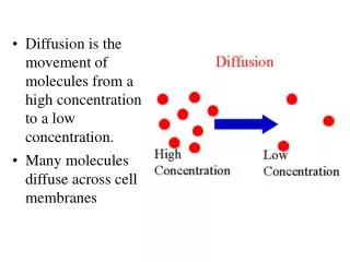
This activity focuses on diffusion experiments using Methylene Blue and Potassium Permanganate to study molecular movement. Students will formulate a hypothesis, measure initial and final diameters of the diffusing substances, and explore the proper usage and maintenance of microscopes. The activity includes preparing wet mounts of different cells and examining them at various magnifications. By learning these techniques, students will understand cellular structures and the principles of diffusion while developing practical microscopy skills.

Exploring Diffusion and Microscope Techniques in Cellular Biology Experiments
E N D
Presentation Transcript
Activity 1 Diffusion experiment Methylene blue MW 320g/mole Potassium permagnate MW 150 g/mole Form Hypothesis T0 initial diameter T60 final diameter Room temp Fridge
Storing The Microscope • Return the lowest power objective in place • Wrap the cord around the base • Return dustcover
Cleaning the Microscope • Use lens paper on all glass parts of the microscope. • Clean oil immersion lens with chemicals provided by your instructor
Parts of the Microscope • Ocular • Body tube • Nosepiece • Objective lenses (10, 40, 100x) • Arm • Course adjustment knob • Fine adjustment knob • Stage • Stage clips • Aperture • Diaphragm • Light • Base
Microscope Parts • Ocular • Body tube • Stage clip • Revolving nose piece • Objective • Arm • Stage • Diaphragm • Lever to move stage clip • Course adjustment • Fine adjustment • Light source • Base
Using the microscope • Always observe using the LOWEST POWER objective first. • Focus using the COARSE ADJUSTMENT KNOB to bring the object into focus. Bring the object into sharp focus by using the fine adjustment knob. • Focus, and then move to a higher power objective, if needed. • Use only the FINE ADJUSTMENT KNOB when using the HIGHEST (longest) POWER OBJECTIVE. • Keep both eyes open to reduce eyestrain. • Determine total magnification of the object by multiplying the power of the ocular (10x) the power by the power of the objective.
Preparing a slide • Using a pipet or dropper, add a drop of water or another solvent to a clean microscope slide. Then, place the specimen in the water. • Place the edge of a coverslip on the slide so that it touches the edge of the water. • Slowly lower the coverslip to prevent the formation of air bubbles.
Oil Immersion Lens 100x 40x
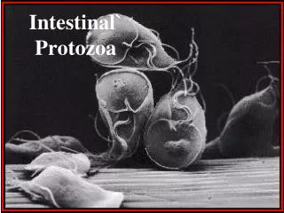
Activity 2 • Prepare wet mount • Cheek cell • Elodea cell • Onion cell • Look at specimen under high power and draw what you see. • Use proper clean-up technique
Activity 3 • Prepare wet mount for dissecting scope • Pond water • Look at specimen under high power and draw what you see. • Use proper clean-up technique
Microscope Quiz • Which objective uses oil? • If your ocular is 10x and your objective is 40x what is the total magnification of your image? • Why do you need a cover slip and how do you avoid an air bubble? • What is the difference between a light microscope and a dissecting microscope? 1.
Instructor’s Notes: • Materials & Equipment • 5 dissecting scopes • 5 light microscopes • Cover slips • Slides (box) • Droppers • Water in dropper • Oil and cleaner for oil immersion lens • Lens paper • Onion • Elodea • Toothpicks (box) • Sharps and biohazard bag • disinfectant • 5 Rulers • 10 agar plates plus a few extra • Methylene blue • Potassium permagnate • Razor blade to cut plants • Pondwater • Use of fridge