Download
1 / 44
460 likes | 482 Vues
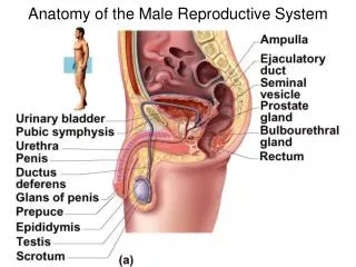
Anatomy of the Male Reproductive System. Scrotum. pouch of skin that hangs at root of penis and contains the testes superficial fascia divides scrotum into right & left halves provides temperature about 3 C below body temperature for proper sperm production. Testes.

E N D
Scrotum • pouch of skin that hangs at root of penis and contains the testes • superficial fascia divides scrotum into right & left halves • provides temperature about 3C below body temperature for proper sperm production
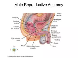
Testes • seminiferous tubules: sperm factories • series of tubes carries sperm from testes to epididymis • interstitial cells or Leydig cells: produce androgens, especially testosterone • testicular arteries & veins: provide blood & help maintain temperature
The Duct System transports sperm from body
Epididymis • stores immature sperm temporarily • takes about 20 days for sperm to mature
Ductus Deferens • Muscle contractions propel live sperm to urethra • during ejaculation smooth muscle contractions rapidly squeezes sperm forward • cut during a vasectomy for birth control
Urethra • conveys urine & semen to tip of penis
Seminal Vesicles • located at base of bladder • produces 60% of fluid in semen • yellowish thick fluid • fructose, ascorbic acid, amino acids, & prostaglandins • sperm & seminal fluid mix in ejaculatory duct
Prostate Gland • 33% of semen volume • milky alkaline fluid that activates sperm
Bulbourethral Glands • Cowper’s glands • produce thick, clear mucus • released prior to ejaculation • neutralize traces of acidic urine & lubricant during intercourse
Semen • mixture of sperm & secretions • transport medium, nutrients, & chemicals that protect & facilitate movement of sperm • hormone relaxin enhance sperm motility • basic pH 7.2 - 7.6 neutralizes acidic environment of vagina
2 - 6 ml released during ejaculation • 50 - 100 million sperm in each ml
Spermatogenesis • sequence of events in seminiferous tubules of testes that leads to production of male gametes or sperm • healthy male produces several hundred million sperm per day
Effects of Testosterone • at puberty testosterone prompts spermatogenesis • causes reproductive organs to grow & assume adult functions • as adult normal levels of testosterone are required to maintain normal structure & function of reproductive organ
Erection • erectile tissue of penis, corpora cavernosa becomes engorged with blood • parasympathetic nerve fibers stimulate arterioles to dilate, increasing blood flow • blood flow is cut off trapping blood causing penis to stiffen and become elongated
Ejaculation • propulsion of semen from male duct system • reproductive ducts & accessory glands contract peristaltically emptying their contents into urethra • bladder sphincter muscle constricts preventing expulsion of urine
bulbospongiosus muscles of penis undergo rapid series of contractions propelling semen from the urethra
Ovaries • inside are many tiny saclike structures called ovarian follicles • each month in adult women one mature follicle ejects its oocyte called ovulation • changes into structure called corpus luteum
Uterine Tubes • fallopian tubes • provide site where fertilization can occur • little or no contact with ovary • fimbriae become active close to ovulation, they create currents in peritoneal fluid & usually carry oocyte into uterine tube
oocyte is carried toward uterus by peristalsis & rhythmic beating of cilia
Uterus • size & shape of pear • hollow, thick-walled organ that functions to receive, retain, & nourish a fertilized egg and developing baby
Uterine Wall • 3 layers • perimetrium: outermost serous layer • myometrium: thick layer of smooth muscles, plays active role in childbirth • endometrium: simple columnar epithelium anchored by thick connective tissue • highly vascular
Vagina • thin-walled fibromuscular tube • birth canal • receives penis & semen during intercourse • pH 3.5 - 4.0 to reduce possibility of infection
External Genitalia • vulva • mons pubis: fatty, rounded area overlying pubic symphysis • labia majora: two elongated, hair-covered fatty skin folds • labia minora: two thin hair-free folds covered with mucosa & sebaceous glands
vestibular glands: by vaginal opening, release mucus for moisture & lubrication during intercourse • clitoris: protruding structure, composed of erectile tissue • becomes swollen during sexual arousal
Mammary Glands • present in both sexes, become functional only in females • areola: pigmented area that surrounds nipple • alveolar glands: produce milk when woman is lactating
lactiferous ducts: carry milk to outside of body • lactiferous sinus or ampulla: sinus where milk accumulates during lactation
Oogenesis • process in which eggs are produced • supply of eggs that female releases is determined by the time of birth • from puberty to about 50 • one ovulation each month • Only 400 - 500 oocytes of potential 700,000 are released during lifetime
Ovarian Cycle • 3 phases • typical cycle lasts 28 days
Follicular Phase • period of follicle growth • days 1 - 10
Ovulatory Phase • days 11 - 14 • ovary wall at site of ballooning ruptures & expels oocyte into peritoneal cavity • 1 - 2% of ovulations more than one oocyte is released, which could result in multiple births
Luteal Phase • days 14 - 28 • oocyte increases in size & now called corpus luteum • begins to secrete progesterone & some estrogen
Hormonal Regulation of Ovarian Cycle • at puberty hypothalamus releases GnRH (Gonadotropin-releasing Hormone) • stimulates release of FSH (follicular-stimulating hormone) & LH (luteinizing hormone) • stimulate growth of follicle
estrogen & progesterone release cause negative feedback or inhibitory effect on release of LH & FSH from anterior pituitary
Uterine (Menstrual) Cycle • 3 stages
Menstrual Phase • thick functional layer of uterine endometrium becomes detached • bleeding for 3 - 5 days • passes through vagina • menstrual flow • 50 - 150 ml of blood lost
Proliferative Phase • days 6 - 14 • estrogen causes endometrium repair • mucosa becomes velvety, thick, & well vascularized • cervical mucus thins to form channels that aid movement of sperm into uterus • ovulation occurs
Secretory Phase • days 15 - 28 • increased level of progesterone • uterus ready for implantation of embryo • cervical plug forms blocks sperm & keeps uterus “private” if embryo implants • no fertilization endometrial cells die