Download
1 / 24
260 likes | 511 Vues
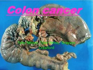
Immunohistochemistry Involving the Use of Certain Antibodies and Colon Cancer. Presenter: Ms. Candice Carr Mentor: Michael Wargovich, Ph. D Location: South Carolina Cancer Center Program: CRTP/ USC School of Medicine & Claflin University. What is Immunohistochemistry?.

E N D
Immunohistochemistry Involving the Use of Certain Antibodies and Colon Cancer Presenter: Ms. Candice Carr Mentor: Michael Wargovich, Ph. D Location: South Carolina Cancer Center Program: CRTP/ USC School of Medicine & Claflin University
What is Immunohistochemistry? • A technique for identifying cellular or tissue constituents (antigens) by means of antigen-antibody interactions, the site of antibody binding being identified either by direct labeling of the antibody, or by use of a secondary labeling method. • Demonstration of specific antigens in tissues by the use of markers that are either fluorescent dyes or enzymes.
Background of Immunohistochemistry • Primary antibody binds to specific antigen • Antibody-antigen complex is bound by a secondary, enzyme-conjugated, antibody. Note: To amplify the staining, we use a complex of peroxidase-antiperoxidase (PAP), avidin-biotin-peroxidase (ABC) or avidin-biotin alkaline phosphatase. 3. In the presence of substrate and chromogen, the enzyme forms a colored deposit at the sites of antibody-antigen binding. The color of the deposit depends on the chromogen used.
Hisotechniques • Tissue Processing a) dehydration b) clearing • Tissue Sectioning • Deparaffinizing • Epitope Retrieval • Immunostaining • Counterstaining • Coverslipping
Dehydration The water from the tissue must be removed by dehydration. This is usually done with a series of alcohol: 70% to 95% to 100%. Clearing Consists of removal of the dehydrant with a substrate. The commonest clearing agent is xylene. Hisotechniques1. Tissue Processing Embedding A technician must pick the tissue out of the cassette and pour molten paraffin over it. The tissue must be aligned and laying flat in the block of paraffin so that the crypts will be showing and not the lumen.
Hisotechinques2. Tissue Sectioning • Once the tissue have been embedded, it must be cut into sections. This is done with a microtome. The sections are placed carefully in a water bath. Retrieve sections from water bath and place on slides. Put slides in slide tray and placed in oven at 600C for an hour to dry.
Hisotechniques3. Deparaffinizing The embedding process must be reversed in order to get the paraffin wax out of the tissue and allow water soluble dyes to penetrate the sections. This is done by running them through xylenes (or substitutes) to alcohols to water. Note: No stains can be done on tissues containing paraffin.
Hisotechniques4. Epitope Retrieval • Pre-heat steamer • Heat contained Citrate Buffer in microwave until it reaches 980C • Place deparaffinized slides in the heated contained solution and put in steamer • Incubate for 20 minutes • Allow slides to cool for 20 minutes • Rinse slides with distilled water • Put slides in TBS Buffer with tween for 10 minutes
Hisotechniques5. Immunostaining The staining process makes use of a variety of dyes that have been chosen for their ability to stain various cellular components of tissue. The routine stain is hematoxylin and eosion (H&E). Other stains are referred to as “special stains” because they are used in specific situations according to their diagnostic need. • PCNA (proliferating cell nuclear antigen) • Ki-67 • Beta-Catenin • APC (adenomatous polyposis coli) • Cox-2
Description of a Normal Colon Tissue • Absorptive and secretory epithelium is shaped into crypts. • Lamina propria surrounds crypts. • The mucosa of the colon is characterized by straight crypts with no villi. • Submucosa is the connective tissue layer that allows the mucosa to move during peristalsis. • Muscularis mucosae is the thin layer of smooth muscle at the boundary between mucosa and submucosa. • Clear “bubbles” that appears in crypt epithelium is the mucus in goblet cells. Goblet cells Lamina propria H & E STAINED COLON
PCNA(proliferating cell nuclear antigen) • Description:The proliferating cell nuclear antigen (PCNA) is a protein. The protein has also been identified as the polymerase-associated protein and is synthesized in early G1 and S phases of the cell cycle. In cells fixed with organic solvents, PCNA is seen to be strongly associated in the nuclear regions where DNA synthesis is occurring, whereas in cells fixed with aldehydes the staining is more diffuse but intense and occurs throughout the cell cycle. This is due to the presence of two basic forms of the PCNA protein, a soluble form sensitive to organic fixation and not involved in replication, and a second form that is insoluble and is associated with ongoing DNA synthesis. • Information about PCNA • Control: tonsil • In tonsil you find appropriate staining for PCNA in the lobes of the tonsil positive control • In colon you find positive staining in the crypts of the colon. • Best fixative for staining PCNA is formalin-fixed, paraffin embedded sections. • Detection kit used when staining PCNA is the LSAB2 kit consisting of: H202, link, HRP-Streptavidin, DAB, antibody-PCNA, and the negative reagent • Stains the nucleus
Slides Stained with PCNA Immunohistochemistry. A: Representative sections from BrdU-injected heterozygous and guanylin null mice were visualized in ileum and distal colon at 1 hour (ileum) and 24 hours (distal colon). B: Representative sections from PCNA-stained ileum and distal colon of heterozygous and guanylin null mice. Bars, 100 µm are shown.
Description:The assessment of cell proliferation by the detection of Ki67 antigen in neoplastic cell populations has been shown to be of prognostic value. The polyclonal antibody (NCL-Ki67p) labels Ki67 antigen in the granular components of the nucleolus during late G1, S, G2 and M phases. There is a strong correlation between low or high Ki67 index and low or high grade histopathology of neoplasms. Information about Ki-67 Control: tonsil In tonsil you find appropriate staining found in the lobes of the tonsil You will find Ki-67 staining in the crypts of the colon Best fixative for staining Ki-67 is formalin-fixed, paraffin embedded sections Detection kit used for staining Ki-67 is the LSAB2 kit consisting of: H202, link, HRP-Streptavidin, DAB, antibody-Ki-67, and the negative reagent Ki-67 stains the nucleus Ki-67 Antibody
Slides Stained with Ki-67 Lower numbers of Ki-67 positive cells (left) and a high percentage of Ki-67 stained cells. Note the prominent nucleolar positivity in tumor cells (right), x100
Beta-Catenin Antibody • Description:The catenins (α, β, γ, δ) are ubiquitously expressed, cytoplasmic proteins that associate with E-cadherin at cellular junctions. α-catenin also associates with P-cadherin and N-cadherin. β-catenin co-immunoprecipitates with APC. γ-catenin associates with both N-cadherin and E-cadherin and is a major component of desmosomes, where it is found complexed to desmoglein. δ-catenin interacts with Presenilin 1. A hemizygous deletion of the gene encoding δ-catenin correlates with mental retardation in cri-du-chat syndrome. Armadillo Repeat gene deleted in Velo-Cardio-Facial syndrome (ARVCF) is also a member of the catenin family that may function as a nuclear protein. • Information about Beta-Catenin • Control: colon • Β-catenin mostly stains the membrane/cytoplasm in normal tissue • β-catenin stains the nucleus when there is cancerous tumors in the the tissue • Best fixative for staining β-catenin is formalin-fixed, paraffin embedded sections • The LSAB2 kit is used when staining with β-catenin consisting of: H202, link, HRP-Streptavidin, DAB, antibody-β-catenin, and the negative reagent • β-catenin can stain both the nucleus and the membrane.
Slides Stained with Beta-Catenin ß-Catenin and phospho-ß-catenin expression on representative colorectal histo-spots on a TMA. Tumors showed either predominantly membranous staining (A) or predominantly cytoplasmic and nuclear staining with ß-catenin (B). With phospho-ß-catenin, there was either no staining (C) or nuclear staining (D). Figures are x100, and insets are x400.
APC(adenomatous polyposis coli) • Description: APC protein plays a major role in colon carcinogenesis because it functions as a negative regulator of cytosolicβ-cateninprotein expression. APC protein is present in a multi-protein complex with β-catenin and two other proteins, axin and glycogen synthase kinase. With in this multi-protein complex, glycogen synthase kinase phosphorylates β-Catenin, leading to its dissociation from the complex and degradation, which occurs via a ubiquitin-dependent proteasomal pathway. • Information about APC • Control: colon • The complete function of the APC gene is not known • It is shown that APC interacts with β-catenin in a multi-protein complex to regulate the level of expression of β-catenin • Loss of the APC gene is required for adenoma formation • Almost all of the mutations found in the APC gene are insertions and deletions mutations that leads to a stop codon and result in the production of a truncated protein that has lost the C-terminal end. • APC is used to see how advance the cell cycle is in the crypts of the colon.
Slides Stained with APC Loss of full-length APC protein in tumors from Apc1638N mice. Specimens of tumor from the small intestines of 65-day-old animals were formalin fixed, embedded in paraffin, and sectioned at 5 µm. Sections were stained with antibody to the COOH terminus of APC protein. As a control, normal small intestinal mucosa from the wild-type littermates of the Apc1638N mouse was stained and found to exhibit cytoplasmic stain for full-length APC. A, small intestinal tumor from a Apc1638N mouse showing a loss of full-length APC protein (x40); B, normal small intestinal mucosa in a Apc1638N mouse showing APC protein staining.
Cox-2 Antibody (mouse) • Information about Cox-2 (mouse) • Control: breast cancer • The APC protein is found in the cytoplasm ofnormal colonic epithelial cells. • Best fixative for Cox-2 (mouse) is formalin-fixed, paraffin embedded sections • The envision+ kit is used when staining Cox-2 (mouse) which consists of: H202, polymer (BPB), pEnV+HRP-Streptavidin, DAB+, antibody-Cox-2 (mouse), the negative reagent. • AOM-induced mouse colon tumors do not express full-length APC protein • Evidence in human colorectal cancer andMIN mice implicate the tumor suppressor gene, APC, in the causation ofcolorectal carcinogenesis • Description: COX-2(Cyclooxygenase-2) is an inducible enzyme. It is involved in the response of cells to growth factors, tumor promoters, and cytokines that induce its expression. Given its role in synthesizing prostaglandins, COX-2 is therefore of interest in studying immune response regulation. COX-2 is induced by a wide variety of stimuli and was initially identified as immediate-early growth response gene. In addition, COX-2 expression markedly increased in 85-90% of human colorectal adenocarcinoma whereas COX-1 levels remain unchanged.
Slides Stained with Cox-2 (mouse) Immunolabeling for COX-2 in 3-µm paraffin-embedded sections of inner retina from C57BL/6 mice with retinopathy of prematurity. Counterstain, hematoxylin. (A) Normal untreated. (B) Untreated ROP. (C) Normal treated with rofecoxib. (D) ROP treated with rofecoxib. Intense COX-2 immunolabeling was observed in ganglion cells (single arrow) and blood vessels (double arrows). Magnification, x250.
Hisotechniques6. Counterstaining • Slides are placed in stainer • Then dipped in 25% Gill’s Hematoxylin Solution (blue), which is mostly used in my lab to give clear and sharp nucleus staining with little background. • Rinsed with distilled water • Dehydrated through 95% ethanol, then through 100% ethanol • Cleared in xylene • Other counterstains include: • Mayer’s Hematoxylin Solution (blue) • Nuclear Fast Red Solution (red) • Methyl Green Solution (green) • PI Counterstain Solution (fluorescent red) • DAPI Counterstain Solution (fluorescent blue)
Hisotechniques7. Coverslipping • After the slides have been counterstained they can coverslipped with a permanent mounting medium manually or by using a coverslip machine. This is done to preserve the section on the slide for future purposes.
Future Direction • To finish helping Dr. Mike write his article on Chemoprevention of Colorectal Cancer in F344 Rats by Rofecoxib • Stained Cox-2 using the human tissue • To learn or found out more about the function of the APC gene and its connection with colon cancer • Actually work with mice in an experiment
Dr. Mike Wargovich South Carolina Cancer Center SCCC Histology Core ~ Ms. Valerie ~ Ms. Sharon Lab 4 & 7 ~Suresh Volate ~Ala Issa ~Cindy Woods ~Scott Hudson ~Hyllaerye Ford Dr. Omar Bagasra Dr. Kim Creek Claflin University USC School of Medicine Acknowledgements