RESULTS
METHODS. Step 1: Design Molecular Probes. A B. A B C D. Karenia brevis, CCMP 718, 100% Karenia brevis, CCMP 718, 50% Karenia brevis, CCMP 718, 25% Karenia brevis , TX-sp3 Karenia brevis , NOAA-1 Karenia mikimotoi, NOAA-2

RESULTS
E N D
Presentation Transcript
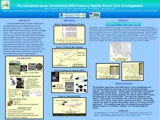
METHODS Step 1: Design Molecular Probes A B A B C D Karenia brevis, CCMP 718, 100% Karenia brevis, CCMP 718, 50% Karenia brevis, CCMP 718, 25% Karenia brevis, TX-sp3 Karenia brevis, NOAA-1 Karenia mikimotoi, NOAA-2 negative PCR control blank Karenia brevis, CCMP 718 Karenia brevis, TX-sp3 Karenia brevis, NOAA-1 Karenia mikimotoi, NOAA-2 negative PCR control blank 5’-CTCATGGTGGCGGCTGG-3’ 1) Henderson Creek 2) Marco Pass 3) 951 boat ramp 4) Caxambas Pass 5) Goodland 6) Blackwater River 7) Faka Union Bay 8) Fakahatchee Bay design probes to be species-specific and to work at a single temperature 1 3 2 Step 2: Produce Microplates 5 6 4 7 8 5’- CTCATGGTGGCGGCTGG -3’ probe + = T-tailed probe to raise probe off plate surface TTTTTTTTTTT Fig. 3. Brevis Probe: 2 bp specificity needed to distinguish K. brevis from K. mikimotoi poly-t’s The microplate assay: Immobilized DNA Probes to Rapidly Detect Toxic Dinoflagellates Kelly D. Goodwin*, Sara A. Cotton+, Gloria Scorzetti**, Traci Kiesling **, and Jack W. Fell** *NOAA Atlantic Oceanographic and Meteorological Laboratories, +Coop. Inst. Marine & Atmos. Studies & **Marine Biology and FisheriesRosenstiel School of Marine and Atmospheric Science, University of Miami BACKGROUND covalent chemistry Harmful Algae and = immobilize probe to wells Fig. 2 Karenia Probe: Detects K. brevis and K. mikimotoi finished microplate Fig. 1. Sampling sites in the Rookery Bay NERR Step 3: Extract & Amplify DNA Sewage Contamination Impact Coastal Water A) filter water sample Water Quality Assays Need Improvement B) extract genomic DNA • Harmful Algae: • require extensive microscopic expertise • hard to distinguish closely related species • samples fragile & hard to preserve C) PCR with biotin-labeled universal primers • Fecal Contamination: • labor intensive • take too long • measure indicators vs. pathogens Step 4: Hybridize DNA Molecular-Based Assays: Blue +Stop Solution = Yellow Enzyme Substrate • Sensitive • Specific • Microscope & Culture Independent Streptavidin-POD Target DNA Biotin Probe Development Goals: • aid managers and decision makers • aid ecological research • provide early and accurate detection • distinguish between human and animal waste • fast, convenient, economical Microplate Well yellow color identifies presence of organism ABSTRACT RESULTS A DNA hybridization assay in microtiter plate format was adapted to detect toxic dinoflagellates and fecal bacteria in coastal waters. The assay provided species-specific identification and simultaneous detection of multiple targets. The assay detected K. brevis in coastal waters collected from the Rookery Bay National Estuarine Research Reserve (NERR). Results were verified by species-specific PCR and sequence analysis. The presence/absence of K. brevis was consistent with microscopic observation. The assay yielded quick colorimetric results, employed a single hybridization temperature, and conserved the amount of genomic DNA utilized by using one set of PCR primers. The microplate assay provides a useful tool to quickly screen large sample sets for multiple target organisms. Detection of Toxic Dinoflagellates The microplate assay provided identification of several toxic dinoflagellates, including Karenia brevis, the organism responsible for red tide in the Rookery Bay NERR (Fig. 1). The assay was evaluated with 110 environmental samples and was consistent with species-specific PCR, sequencing, and microscopic observation. Target sensitivity for this study was a negative result consistent with a report of “not present” provided by a regional monitoring program. With HPLC purified probes, this objective was met in every case tested. Assay sensitivity allowed detection of K. brevis when it was “present” in the water (<1000 cells/L), as defined by the Florida Marine Research Institute (FMRI). Simultaneous and species-specific detection of multiple targets was demonstrated with K. brevis and Amphidinium carterae and was achieved with a single hybridization condition and one set of PCR primers. Immobilized “Karenia” probe detected both K. brevis and Karenia mikimotoi (Fig. 2). The “Brevis” probe distinguished between these closely related species (Fig. 3). CONCLUSTIONS The microplate assay allows rapid identification of toxic dinoflagellates and fecal bacteria without the microscopic expertise and culturing normally required. The assay can detect multiple species simultaneously and distinguish between closely related species. The technique does not require expensive instrumentation and has several advantages over species-specific PCR or cloning and sequencing of total extracted DNA. The assay gives immediate visual results, is more specific and convenient than a series of species-specific PCR reactions, and is faster, easier and less expensive than cloning. The technique conserves the amount of genomic DNA utilized, which can be critical to certain applications4. The microplate assay offers the sensitivity and specificity of molecular analysis in a convenient, adaptable, and relatively inexpensive format. REFERENCES 1based on Regnault et al. Res. Microbiol. 2000, V151, pp. 521-533, 2based on Loge et al. Water Env. Res. 1999, 71:76-83; 3based on Franks et al. Appl. Environ. Microbiol. 1998, 64: 3336-3345; 4Kiesling, T.L., Wilkinson, E., Rabalais, J., Ortner, P.B., McCabe, M.M. and Fell, J.W., Mar. Biotechnol. 2002, 4:30-39.