Download
1 / 46
460 likes | 645 Vues
Protein Synthesis Dr. M. Jawad Hassan. WHAT IS TRANSLATION?. The process by which genetic information contained within the order of nucleotides in messenger RNA (mRNA) is used to generate the linear sequences of amino acids in proteins, is called Translation .

E N D
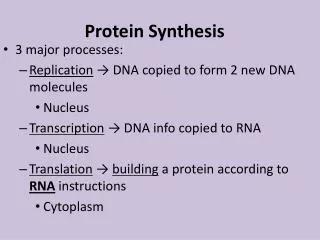
WHAT IS TRANSLATION? • The process by which genetic information contained within the order of nucleotides in messenger RNA (mRNA) is used to generate the linear sequences of amino acids in proteins, is called Translation. • Translation is among the most highly conserved processes across all organisms & among the most energetically costly for the cell.
TRANSLATIONAL MACHINERY • The machinery responsible for translating the language of messenger RNAs into the language of proteins is composed of four primary components: • mRNA • tRNA • Aminoacyl tRNA synthetases • Ribosomes • Amino acids
MESSENGER RNA • The messenger RNA provides the information that must be interpreted by the translation machinery, & is the template for translation. • The protein-coding region of mRNA consists of an ordered series of three-nucleotide-long units called codons that specify the order of amino acids. • The protein coding region(s) of each mRNA is composed of a contiguous, non-overlapping string of codons called an open-reading frame commonly known as an ORF. • Each ORF specifies a single protein & starts & ends at internal sites within the mRNA. That is, the ends of an ORF are distinct from the ends of the mRNA.
Translation starts at the 5’ end of the open-reading frame and proceeds one codon at a time to the 3’ end. • The first & last codons of an ORF are known as the start & stop codons respectively. • In bacteria, the start codon is usually 5-’AUG-3’ but 5’-GUG3’ & sometimes even 5’-UUG-3’ are also used. • Eukaryotic cells always use 5’-AUG-3’ as the start codon. • This codon has two important functions. • First, it specifies the first aa to be incorporated into the growing polypeptide chain. • Second, it defines the reading frame for all subsequent codons.
Stop codons, of which there are three (5’-UAG-3’, 5’-UGA-3’, & 5’-UAA-3’), define the end of the open-reading frame & signal termination of polypeptide synthesis. • It must be noted that genetic code is degenerate that is each amino acid is usually specified by more than one codon. • Eukaryotic mRNA almost always contain a single ORF. In contrast, prokaryotic mRNA frequently contain two or more ORFs& hence can encode multiple polypeptide chains. • Messenger RNAs containing multiple ORFs are known as polycistronic mRNAs. • Messenger RNAs encoding a single ORF are known as monocistronic mRNAs.
To facilitate binding by a ribosome, many prokaryotic open-reading frames contain a short sequence upstream (on the 5’ side) of the start codon called the ribosome binding site (RBS). • This element is also called Shine-Dalgarno sequence. TRANSFER RNA • Transfer RNA molecules act as adaptors between codons & the amino acids they specify. • They have anticodons which dictate the amino acids that are responsible for incorporating into the growing polypeptide chain.
tRNA molecules exhibit a characteristic pattern of single-stranded and double-stranded regions (secondary structure) that can be illustrated as a cloverleaf. • The actual three-dimensional configuration of this adaptor molecule by X-ray crystallography reveals an L-shaped tertiary structure.
tRNA molecules to which an amino acid is attached are said to be charged, & tRNAs that lack an amino acid are said to beuncharged. • Charging requires an acyl linkage between the carboxyl group of the amino acid & the 2’- or 3’-hydroxyl group of the adenosine nucleotide that protrudes from the acceptor stem.
AMINOACYL tRNA SYNTHETASE • All aminoacyl tRNA synthetases attach an amino acid to a tRNA in two enzymatic steps. • Step one is adenylylationin which the amino acid reacts with ATP to become adenylylated with the concomitant release of pyrophosphate. • As a result of adenylylation, the amino acid is attached to adenylic acid via a high-energy ester bond in which carbonyl group of the amino acid is joined to the phosphoryl group of AMP.
Step twois tRNA charging in which the adenylylated aa which remains tightly bound to the sythetase, reacts with tRNA. • This reaction results in the transfer of the amino acid to the 3’ end of the tRNA via the 2’- or 3’- hydroxyl & the concomitant release of AMP.
There are two classes of aminoacyl tRNA synthetases: CLASS I ENZYMES attach the amino acid to the 2’OH of the tRNA & are generally monomeric. CLASS II ENZYMES attach the amino acid to the 3’OH of the tRNA & are typically dimeric or tetrameric. One common mechanism to increase the fidelity of an aminoacyl tRNA synthetase is to proofread the products of the charging reaction as seen for DNA polymerase.
RIBOSOMES • The ribosome is the macromolecular machine that directs the synthesis of proteins. • The ribosome is composed of two subassemblies of RNA & protein known as large and small subunits.
The large subunit contains the peptidyl transferase center, which is responsible for the formation of peptide bonds. • The small subunit contains the decoding center in which charged tRNA read or “decode” the codon units of the mRNA. • The small & large subunits of ribosome associate & dissociate with each other after each round of protein synthesis. This is called ribosome cycle. • Each mRNA can be translated simultaneously by multiple ribosomes. An mRNA bearing multiple ribosomes is known as a polyribosomeor a polysome.
The ribosome catalyzes a single chemical reaction: the formation of peptide bond. • This reaction occurs b/w the aa residue at the carboxy-terminal end of the growing polypeptide & the incoming aa to be added to the chain. • To carry out peptidyl transferase reaction, the ribosome must be able to bind at least two tRNAs simultaneously. • In fact, the ribosome contains three tRNA binding sites, theA, P, and E sites.
The A site is the binding site for the Aminoacylated-tRNA which is attached at its 3’ end to the carboxyl group of the amino acid. • The P site is the binding site for the Peptidyl-tRNA which is attached at its 3’ end to the carboxyl-terminus of the growing polypeptide chain. • The E site is the binding site for the tRNA that is released after the growing polypeptide chain has been transferred to the aminoacyl-tRNA (Eis for exit).
PHASES OF TRANSLATION • There are three stages of translation namely: • Initiation of the synthesis of a new polypeptide chain • Elongation of the growing polypeptide • 3. Termination of polypeptide synthesis
1- INITIATION • For translation to be successfully initiated, three events must occur. • First, the ribosome must be recruited to the mRNA. • Second, a charged tRNA must be placed into the P site of the ribosome. • Third, the ribosome must be precisely positioned over the start codon.
i) PROKARYOTIC TRANSLATIONAL INITIATION • For prokaryotes the association of the small subunit with the mRNA is mediated by base-pairing interactions between the ribosomes binding site & 16S rRNA. • Typically charged tRNA enter the ribosome in the A site and only reach Psite after a round of peptide bond synthesis. • Duringinitiation, however, a charged tRNA enters the P site directly. • This event requires a special tRNA known as the initiator tRNA, which base-pairs with the start codon-usually AUGor GUG.
AUG and GUG have a different meaning when they occur within an open-reading frame, where they are read by tRNA for methionine and valine respectively. • Neither methionine nor valine is attached to the initiator tRNA. Instead, it is charged with a modified form of methionine (N-formyl methionine) that has a formyl group attached to its amino group. • The charged initiator RNA is referred to as fMet-tRNAifMet. • An enzyme known as a deformylase removes the formyl group from the amino terminus during or after the synthesis of polypeptide chain.
The initiation of prokaryotic translation commences with the small subunit and is catalysed by three translation initiation factors namely IF1, IF2, and IF3. • With all three initiation factors bound, the small subunit is prepared to bind to the mRNA & the initiator tRNA. These two RNAs can bind in either order & independently of each other. • Of the three potential tRNA binding sites on the small subunit, only the P site is capable of binding a tRNA in the presence of the initiation factors. • The last step of prokaryotic initiation involves the association of the large subunit to create the 70S initiation complex.
The net result of initiation is the formation of an intact (70S) ribosome assembled at the start site of the mRNA with fMet-tRNAifMet in the P site & an empty A site. The ribosome-mRNA complex is now poised to accept a charged tRNA into the A site & commence polypeptide synthesis.
ii) EUKARYOTIC TRANSLATIONAL INITIATION • Initiation of translation in eukaryotes is similar to prokaryotic initiation in many ways but eukaryotes use a fundamentally distinct method to recognize the mRNA & the start codon. • More than 30 different auxiliary factors are involved in eukaryotic translational initiation. In eukaryotes, the small subunit is already associated with an initiator tRNA when it is recruited to the capped 5’ end of the mRNA. • It then “scans” along the mRNA in a 5’ 3’ direction untill it reaches the first 5’-AUG-3’ in the correct context.
As the eukaryotic ribosome completes a cycle of translation, it dissociates into free large and small subunits through the action of factors . • These factors are called eIF3, eIF1A, analogous to the prokaryotic initiation factors IF3 & IF1 respectively. • Two GTP-binding proteins, eIF2 & eIF5B, mediate the recruitment of the charged initiator tRNA. • For eukaryotes this tRNA is charged with methionine and is refered to as Met-tRNAiMet. • The eukaryotic analog ofIF2-GTP (prokaryotic factor)is eIF5B-GTP.
Once assembled at the 5’ end of the mRNA, the small subunit and its associated factors move along the mRNA in a 5’ 3’ direction in an ATP-dependent process that is driven by the eIF4F-associated RNA helicase. • During this movement, the small subunit “scans” the mRNA for the start codon. • The start codon is recognized through base pairing between the anticodon of the initiator tRNA & the start codon of mRNA. • Correct base-pairing triggers the release of eIF2 & eIF3 allows the large subunit to bind to the small subunit. • The binding of the large subunit leads to the release of the remaining initiation factors by stimulating GTP hydrolysis by eIF5B.
As a result of these events, the Met-tRNAiMet is placed in the P site of the resulting 80S initiation complex. • With the start codon & Met-tRNAiMet,placed in the P site, the eukaryotic ribosome is now poised to accept a charged tRNA into its A site & to carry out the formation of first peptide bond.
2- ELONGATION • Once the ribosome is assembled with the charged initiator tRNA in the P site, polypeptide synthesis can begin. • There are three key events that must occur for the correct addition of each aa. • First, the correct aminoacyl-tRNA is loaded into the A site of the ribosome as dictated by the A-site codon. • Second, a peptide bond is formed b/w the aminoacyl-tRNA in the A site & the peptide chain that is attached to the peptidyl-tRNA in the P site.
This peptidyl transferase reaction results in the transfer of the growing polypeptide from the tRNA in the P site to the aa moiety of the charged tRNA in the A site. • Third, the resulting peptidyl-tRNA in the A site & its associated codonmust be translocated to the P site so that the ribosome is poised for another cycle of codon recognition & peptide bond formation. • Two auxiliary proteins known as elongation factors control these events. Bothof these factors use the energy of GTPbinding & hydrolysis to enhance the rate & accuracy of ribosome function.
EF-Tucan only bind to an aminoacyl-tRNAwhen it is associated with GTP. • When EF-Tu hydrolyzes its bound GTP, any associated aminoacyl-tRNA is released. • The trigger that activates the EF-Tu GTPase is the same domain on the larger subunit of the ribosome that activates the IF2 GTPase or eIF5B GTPase when the large subunit joins the initiation complex. • This domain is known as the factor binding center.
Once the correctly charged tRNA has been placed in the A site and has rotated into the peptidyl transferase center, peptide bond formation takes place. • This reaction is catalyzed by RNA as peptidyl transferase center is a ribozyme, that is enzyme composed of RNA. • Once the peptidyl transferase reaction has occurred, the tRNA in the P site is deacetylated & the growing polypeptide chain is linked to thetRNA in the A site.
For a new round of peptide chain elongation to occur, the P site tRNA must move to the E site & the A site tRNA must move to the P site. • At the same time, the mRNA must move by three nucleotides to expose the next codon. • These movements are coordinated within the ribosome & are collectively referred to astranslocation. • Translocation in the large subunit precedes translocation in the small subunit. • The completion of translocation requires the action of a second elongation factor called EF-G.
3-TERMINATION • The ribosome’s cycle of aminoacyl-tRNA binding, peptide bond formation, and translocation continues untill one of the three stop codons enters the Asite. • Stop codons are recognized by proteins calledrelease factors (RFs) that activate the hydrolysis of the polypeptidefrom thepeptidyl-tRNA. • There are two classes of release factors. Class I release factors recognize the stop codons & trigger hydrolysisof the peptide chain from the tRNA in the P site.
Prokaryotes have two class I release factors called RF1 & RF2. • RF1 recognizes the stop codon UAG, & RF2 recognizes the stop codonUGA. • The third stop codon , UAA, is recognized by both RF1and RF2. • In eukaryotic cells there is a single class I release factor called eRF1 that recognizes all three stop codons.