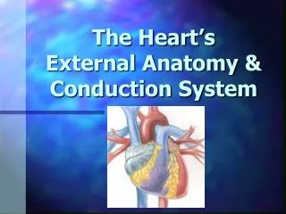
The Heart’s External Anatomy & Conduction System
190 likes | 621 Vues
The Heart’s External Anatomy & Conduction System. Heart at rest Blood flows from large veins into atria Passive flow from atria into ventricles Atria (R & L) contract simultaneously Blood forced into ventricles Ventricles (R & L) contract simultaneously

The Heart’s External Anatomy & Conduction System
E N D
Presentation Transcript
Heart at rest • Blood flows from large veins into atria • Passive flow from atria into ventricles • Atria (R & L) contract simultaneously • Blood forced into ventricles • Ventricles (R & L) contract simultaneously • Atrioventricular valves close “lubb” sound • Blood forced into large arteries • Ventricles relax • Semilunar valves close “dub” sound • Heart at rest
Pericardium • Membrane sac • Surrounds the heart • Protection • Anchors • Contains serous fluid HEART Pericarditis inflammation of the pericardium decreases serous fluid causing painful adhesions interfering with heart movements Pericardium
Heart Wall • Epicardium (outside) – visceral layer of the serous pericardium. • Myocardium (muscle) – cardiac muscle layer forming the bulk of the heart. • Endocardium (within) – endothelial layer of the inner myocardial surface.
Cardiac Muscle • Specialized muscle cells • Involuntary • Striated • Cushioned by endomysium • Joined by intercalated discs • Cardiac cell metabolism • Areobic • Large mitochondria • Organic fuels: fatty acids & glucose • Fatigue resistance
Coronary Arteries Branch off aorta above aortic semilunar valve • Left coronary artery • supplies left atrium and left ventricle • Anterior interventricular artery • supplies both ventricles • Right coronary artery • supplies right ventricle • Posterior interventricular artery • supplies both ventricles
Coronary Veins • Collects wastes from cardiac muscle • Drains into a large sinus on posterior surface of heart called the coronary sinus • Coronary sinus empties into right atrium
The heart beats because of the spread of electrical impulses to the heart muscle, causing it to contract.
Cardiac Conduction System • Cardiac muscle tissue exhibits autorhythmicity = generates its own stimulation. • This is possible because of an internal cardiac conduction system which can initiate and distribute electrical impulses.
Cardiac Conduction System • Comprised of interconnected structures • Sinoatrial node • Atrioventricular node • Atrioventricular Bundle • Bundle Branches • Purkinje Fibres
Sinoatrial (SA) Node • Natural Pacemaker • Upper RA • Neuromyocardial cells • Sympathetic & parasympathetic • Sympathetic ↑HR • Parasympathetic ↓HR
Atrioventricular (AV) Node • Junction of atria and ventricles • Spread of depolarisation - from atrialmyocardium • Delay 0.15 seconds • Time atria to expel blood • Time for ventricular filling • Protection to ventricles • Less autonomic nervous control than SA node • Sympathetic ↑conduction time • Parasympathetic ↓conduction time Atrioventricular node
Depolorization The heart is autorhythmic • Depolarization begins • in sinoatrial (SA) node • Spread through atrial myocardium • Results in myocardial contration of the atria • Delay in atrioventricular (AV) node • To the Bundle of His • AKA atrioventricular bundle
Depolorization The heart is autorhythmic • Separates into 2 main • branches left & right • Located in the interventricular septum • Left bundle – antero-superior division • Right bundle – postero-inferior division • Bundle branches divide - small, dense network of conduction tissue called the Purkinje Fibers • Entire musculature depolarizes quickly
Electrocardiogram Variations in electrical potential radiate from the heart ECG records electrical events in the heart.
P-P = one cardiac cycle • P-Q = time for atrial depolarization • Q-T = time for ventricular depolarization • T-P = time for relaxation • P wave • Depolarization of atria • Followed by contraction • QRS complex • 3 waves (Q, R, & S) • Depolarization of ventricles • Followed by contraction • T wave • Repolarization of ventricles • P-Q interval • Time atria depolarize & remain depolarized • Q-T interval • Time ventricles depolarize & remain depolarized
P T PR QRS SA node Represented on the ECG as P wave AV node conduction is represented on the ECG as the PR Interval The Bundle Branch and purkinjefibredepolarisation constitutes ventricular depolarisation Represented on the ECG as the QRS Atrialrepolarisation occurs within the QRS & therefore is masked Ventricular repolarisation is represented on the ECG as a T wave
1) atrialdepolarization begins 2) atrial depolarization complete (atria contracted) 3) ventriclesbegin to depolarize at apex; atriarepolarize(atria relaxed) 4) ventricular depolarization complete (ventricles contracted) 5) ventriclesbegin torepolarize at apex 6) ventricular repolarization complete(ventricles relaxed)