Download
1 / 31
310 likes | 386 Vues
Explore the radiologic patterns and clinical associations of spinal cord involvement in MS. Detailed findings on lesion characteristics, demographics, clinical presentations, and disease courses. Comprehensive analysis for better understanding and management of MS patients.

E N D
Clinico-Radiological Profile of Spinal Cord Multiple Sclerosis Glenn H. Roberson Bhavik N. Patel Asim K. Bag University of Alabama at Birmingham, Birmingham, AL, USA
Glenn H. Roberson: Involved in clinical trials sponsored by Guerbet LLC & Wyeith Pharmaceuticals Bhavik N. Patel: No disclosure Asim K. Bag: Involved in clinical trials sponsored by ACRIN & Guerbet LLC
Introduction • Multiple sclerosis (MS) has extensive disease burden • MS affects approximately 350,000 individuals in the United States • Typically between the ages of 18 and 45 Medical Clinics of North America 2009;93:451-476
Introduction • Initial MRI diagnosis of MS does not include spinal cord MRI findings • Spinal cord is involved in >90% of MS patients • Asymptomatic cord lesions are found in 30% to 40% of patients • Spinal cord imaging is very important to identify disease progression in time and space Neuroimaging Clinics of North America 2009;19:81-99
Purpose • To identify radiologic pattern of spinal cord involvement in MS • To correlate radiologic findings with clinical symptoms
Materials & Methods: Patients • Retrospective identification of all consecutive patients with abnormal T2 signal in the spinal cord with radiologic concern for MS between 2004 and 2009 • Inclusion criteria • Patients who meet the Revised McDonald MS Diagnostic Criteria were included in this study
Materials & Methods: MRI sequences • Axial • T1 • T2 • STIR • T1+c • Sagittal • T1 • T2 • STIR • T1+c
Materials & Methods: Lesion Characterization • Number of lesions per patient • Involvement pattern of the cord (anterior, posterior, central and diffuse) • Location (cervical, thoracic and lumbar) • Length of lesions • Enhancement pattern
Materials & Methods: Clinical Evaluation • Demography of the patient (age, sex and race) • Clinical presentation • Pattern of disease course
Materials & Methods • Association between lesion location and distribution with symptoms • Association between lesion load and disease course
Results • 544 patients were identified with spinal cord T2 abnormality with radiologic concern for MS • Only 166 patients met the Revised McDonald MS Diagnostic Criteria
Results: Demography • Age range: 17-75 • Male:Female 1:12.9 • More common in Caucasian than African-American (1.84:1)
Results: Clinical presentations • Sensory 42.77% • Motor 37.95 % • Gait 21.68 % • Bladder 12.65 % • No Spinal symptom 12.65 % • Lhermitte 3.01 %
Results: Clinical Course • Relapsing remitting 71.68% • Secondary progressive 24.09% • Primary progressive 0% • Progressive relapsing 0% • Neuromyelitis optica 4.21% Diagram
Results: Lesion loads & disease course • Relapsing-remitting • Average number of lesion 2.20 (range 1 to 7) • Secondary-progressive • Average number of lesion 2.14 (range 1 to 5)
Results: Lesion Characterization 166 patients had total 340 lesions • Location • 46.47% posterior • 27.94% anterior • 22.35% central • 3.23% diffuse • Enhancement • 4.4% • Lesion length • Mean 18.2 mm [range3-108 mm] • Average number of lesions per patient • 2.04
Imaging example Sagittal & Axial T2
Imaging example Sagittal & Axial STIR
Imaging example Sagittal & Axial T2
Imaging example Sagittal & Axial STIR
Imaging example Sagittal & Axial STIR
Imaging example Sagittal T1, T2 & STIR
Imaging example Pre- & post-contrast axial and sagittal T1
Results: Lesion Location • Number of lesions in this bar diagram exceeds 340 as some of the lesions involved more than one segments • Only 7 patients had isolated thoracic spine involvement
Results • No association between lesion location and • Sensory symptoms • Bladder symptoms • Motor symptoms
Results • All patients with posterior column signs, positive Romberg test and gait abnormality had posterior lesions
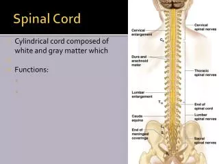
Source: http://en.wikipedia.org/wiki/File:Spinal_cord_tracts_-_English.svg
Limitations • The study is based on retrospective analysis of data • There is a component of selection bias as the study patients were identified from prior MRI
Conclusion • Demography • Age of presentation 17-75 • Predominantly in women (13:1) • Clinical Presentation • Most common presentation is sensory symptoms • Relapsing-remitting is the most common clinical course
Conclusion • Radiologic appearance • Cervical spinal cord is most commonly involved • Posterior spinal cord is involved most commonly • Mean lesion length is 18.2 mm • Enhancement is rare • Clinico-radiologic correlation • Posterior column signs and gait abnormality are associated with posteriorly located lesions • Average number of lesions is similar in relapsing-remitting and secondary progressive MS