
Exploring the Complement System: Pathways and Functions in Immune Defense
E N D
Presentation Transcript
The Complement System By: Dr. Suzan Yousif
Introduction • The complement system consists of a group of serum proteins that act in concert and in an orderly sequence to exert their effect • These proteins are not immunoglobulins and their concentrations in serum do not increase after immunization • Complement activation (fixation) leads to lysis of cells and to the generation of many powerful biologically active substances
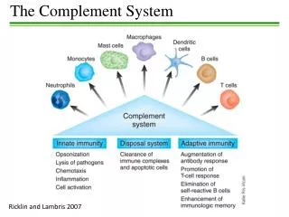
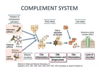
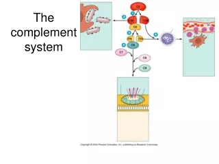
Overview of Complement Classical Pathway Alternative Pathway Lectin Pathway Antibody binds to specific antigen on pathogen surface Clearance of Apoptotic Cells Complement Activation Formation of C3 and C5 convertases Inflammatory response Mannose-binding protein binds pathogen surface Pathogen surface creates environment conducive to complement activation Opsonization & phagocytosis of some pathogens Clearance of Immune complexes Membrane Attack Pathway Cytolysis of some pathogens
Complement Activation Pathways • The Classical Pathway - Ag- Ab complexes • The Alternative Pathway - Aggregated immunoglobulins and microbial products • The Mannan - Binding Lectin Pathway - Microbial products
The Classical Pathway • Activators: Ag – Ab complexes • Antibodies involved: IgG and IgM • Activation in an orderly fashion of nine major protein components; C1 – C9 • Products of activation are enzymes that catalyze the subsequent step
The Classical Pathway • Activation of C1: • C1 consists of C1q (400.000 Daltons), C1r (95000 Daltons), and C1s (85000 Daltons) • Subunits are held together by Calcium ions • C1q is a polymer of 6 identical units • C1q activation requires binding to a c1q- specific receptor on the FC region of at least 2 adjacent molecules of IgG or a single molecule of IgM, a reaction that requires Calcium ions
The Classical Pathway • IgG4, IgA, and IgE do not have complement receptors • Activated C1q activates C1r which in turn activates C1s • Activated C1s has esterolytic and proteolytic properties which acts on C4 splitting it into two fragments; C4a and C4b • C4b complexes with C1s forming an active component that acts on C2 splitting it into C2a and C2b • C2a binds to C4b creating a very active complex called the C3 convertase, where a single molecule can activate hundreds of C3 molecules
The Classical Pathway • C3 is split by C4b2a into C3a and C3b • C3b binds to cells and to C4b2a to generate C5 convertase which splits C5 into C5a and C5b • C5b binds to cells and activates C6 and C7 • The complex C5b67 activates C8 and C9 forming a giant molecule with a molecular weight of 106 Daltonscalled the membrane attack complex (MAC)
The Classical Pathway • C5b6789 bound to cells insert themselves into the cell membrane and produce transmembrane channels allowing ions to pass through • The osmotic equilibrium of the cell is disturbed with rapid influx of water into the cell which swells and lyses
The Alternative (properdin) Pathway • Activators: Bacterial LPS, cell wall of some bacteria, some yeast cells, aggregated IgA, and a factor present in cobra venom • Components: C3 – C9, factor B, factor D, and Properdin • C3b present in trace amounts in serum combines with factor B forming C3bB
Alternative Pathway Figure 16.14
The Mannan Binding Lectin (MBL) • Activators: microorganisms and foreign invaders • Components: C2 – C9, MASP • MBL recognizes carbohydrate structures through its carbohydrate – recognizing domain (CRD) and then it can interact with an enzyme called MBL – activated serine protease (MASP)
Lectin Pathway Figure 16.15
Overview of Complement Classical Pathway Alternative Pathway Lectin Pathway Antibody binds to specific antigen on pathogen surface Clearance of Apoptotic Cells Complement Activation Formation of C3 and C5 convertases Inflammatory response Mannose-binding protein binds pathogen surface Pathogen surface creates environment conducive to complement activation Opsonization & phagocytosis of some pathogens Clearance of Immune complexes Membrane Attack Pathway Cytolysis of some pathogens
Complement functions related to immune defense • Lysis of cells: This is the original function identified and causes hypotonic cell death by making holes. It is not effective against organisms with rigid cell walls such as fungi
Terminal complement components and the formation of the membrane attack complex
The contents of the cell leak out through the MAC pore and the cell dies Before complement After complement treatment
opsonized bacterium Opsonization: Antigen coated with C3b binds to cells bearing complement receptors and if the cell is a phagocyte, the antigen will be phagocytosed.
Inflammation: - Anaphylatoxins: C5a, C3a, and C4a of which C5a is the most potent bind receptors on mast cells and basophils and cause degranulation with the release of pharmacologically active mediators which induce smooth-muscle contraction and increases in vascular permeability. - Chemoattractants: C3a, C5a and C5b67 attract and induce monocytes and neutrophils to adhere to vascular endothelial cells, extravasate through the endothelial lining of the capillaries and migrate to the site of complement activation in the tissue.
Inflammation anaphylotoxins C3a, C4a---increased vascular permeability C5a—chemoattraction C3a, C4a----activation
Immune clearance: Removes immune complexes from the circulation and deposits them in the liver where they are degraded. • Virus neutralization: Complement mediates viral neutralization by facilitating viral aggregation and by coating the viral surface.
Clearance of Apoptotic Cells CR3, CR4 C3b phagocyte • Phagocyte recognizes C3b deposited on the surface of apoptotic cell • Apoptotic cell is ingested and destroyed by phagocyte • This is an important mechanism for clearing self antigens and preventing autoimmune responses • Uptake of apoptotic cell also induces self tolerance, thereby prevents autoimmune response