
Radiographic Anatomy
E N D
Presentation Transcript
Radiographic Anatomy Digestive System
Educational Objectives By the end of this lecture you should be able to: • Identify the anatomical parts of the digestive system on diagrams and radiographs. • Identify the relations between the different parts of the GIT • Explain how to hang GIT radiographs on the view box • State and locate the surface land marks associated with the abdomen.
References 1. Text book of radiographic positioning and related anatomy; by Kenneth L.Bontrager,6th edition. 2. Introduction to Human Anatomy and Physiology: by Eldra Pearl Solomon:W.B.Saunders Company 3. Handbook of Anatomy and physiology for Students of Medical Radiation Technology: Mallett.M:Jaspar Websites http://www6.district125.k12.il.us/science/anatomy/ http://www.innerbody.com/htm/body.html http://www.e-radiography.net/ http://www.getbodysmart.com/index.htm
Digestive System (gastrointestinal; GI tract) • Stomach • Fundus (fluid level seen in erect position) • Body • Pyloric Antrum • Small intestines (small bowel) • Duodenum ("c" shape; bulb) • jejunum • ileum
Digestive System (gastrointestinal; GI tract) • Large intestine (colon) • Caecum (valve; appendix) • Ascending colon • Hepatic flexure • Transverse colon • Splenic flexure • Descending colon • Sigmoid colon (flexure) • Rectum and anal canal • Accessory GI tract organs • Salivary glands • Liver &Gall bladder • Pancreas
Quadrants & Regions of the abdomen • Abdomen divisions • 9 Regions (anatomically) 4 Quadrants (clinically)
Quadrants & Regions of the abdomen MSP: mid-sagittal plane TUP: transumblical plane (L4/5) RLL: right lateral plane LLL : left lateral plane TPP: transpyloric plane (L 1) TTP: transtubercular plane (L 5)
Digestive System (I): Alimentary canal: • 9 m length • Extend from mouth to anus ►Oral cavity ►Pharynx ►Esophagus ►Stomach ►Intestine (small & large) • (II): Accessory organs: • Salivary glands • Pancreas • Liver and biliary system
Pharynx ◙ Levels : from skull base to level of C-6 (13 cm). ◙ 3 parts: • (I): Nasopharynx: • Skull base to the level of soft palate • Anterior: nasal cavity (posterior nares) • Inferior: nasopharyngeal isthmus • Lateral wall: opening of auditory tube • Roof: adenoid • (II): Oropharynx: • Level of soft palate to tip of epiglottis • Anterior: oropharyngeal isthmus • (III): Laryngopharynx: • Tip of epiglottis to level of C-6 • Pyriformfossa
Esophagus ◙ Levels : from C-6 to T-11 (25 cm). Normal points of narrowness: (1) Level of cricoid cartilage; (2) Level of left main bronchus;(3) Passing through the diaphragm. Venous drainage of the lower oesophagus form a point of communication between portal and systemic veins; any obstruction of the portal venous system may lead to oseophagealvarices. ◙ Relations: (3 areas) • (I): In the neck: • Anterior: trachea, thyroid • Posterior: cervical vertebrae • Lateral: common carotid artery • (II): In the thorax: • Anterior: trachea, Lt. main bronchus, Lt. atrium • Posterior: thoracic vertebrae, thoracic duct, descending aorta • Lateral: • Right side: azygous vein, right lung • Left side: • Superior med.: Lt. subclavian artery, aortic arch, Lt. lung • Inferior med.: descending aorta, Lt. lung • (III): In the abdomen: 1-3 cm ; the phrenicampulla lies just above the cardia and may simulate hiatus hernia. The abdominal part is called (submerged segment) and help to prevent reflux from the stomach. Other factors: Acute gastro-oesophagealangle,pressure of right crus of the diaphragm and intrinsic muscles sphincter.
Stomach ◙ Shape: J-shaped, but may varies (volume, position, resp., build) • ◙ 2 Orifices: • Cardiac • Pyloric • ◙ 2 Curvatures: • Lesser • Greater • ◙ 3 Parts: • Fundus (air bubble) • Body • Antrum • ◙ Mucosa: gastric rugae • - Longitudinal: on lesser curvature • - Random (mosaic): elsewhere • ◙ Muscles: • Outer: longitudinal • Inner: circular • Innermost: oblique
Relations of the Stomach • ◙Anterior: • Diaphragm • Left lobe of the liver • Left costal cartilage • Anterior abdominal wall • ◙Posterior (stomach bed): • Diaphragm • Left suprarenal gland • Left kidney • Pancreas • Spleen and splenic artery • Transverse colon and Splenic Flexure. • ◙Stomach lie: The fundus of the stomach is located posterioly while the pyloric antrum is very near to the anterior abdominal wall; so with barium studies(1) In the erect position: Air fluid level seen.(2) Supine: barium fill the fundus while pyloric region is seen in double contrast.(3) Prone: barium fill the pylorus while the fundus is seen in double contrast. • ◙Incisura: is that part of the stomach where there is sudden change in the plan of the stomach from the vertical to the horizontal; it help to show whether the stomach is eutonic, hypertonic or hypotonic according to its level in comparison with the 1st part of the duodenum.
Small intestine ◙ Extension: From pyloric orifice of stomach to ilio-caecal valve ◙ Length: 6 meters (range, 3-10) • ◙3 Parts: • Duodenum • Jejunum • Ileum • ◙ Movements: • Rhythmic • Pendular • Peristaltic
Duodenum ◙C-shaped around the head of pancreas ◙ The shortest ◙ Thewidest ◙ 4 parts: • Duodenal bulb: 2 inches, level of L-1, conical shape • Descending: 3 inches, level of L-2 • Transverse: 4 inches, level of L-3 • Ascending: one inch, level of L-2 • NB: 1. Duodenal bulb (Cap): is a common site of ulcers. It likely seen better in the right anterior oblique. • 2. Descending part: forms a curve around the head of the pancreas; the common bile duct and the pancreatic duct open by a common opening (ampulla of Vater) = {duodenal papilla} : through it. The opening is surrounded by sphincter of Oddi. • 3. During contrast examination; barium reach the duodenal cap after 5minutes, delay emptying more than 15 minutes may be due to obstruction.
Relations of the Duodenum • Duodenal bulb: • Superior and anterior: liver and gall bladder • Inferior: head of pancreas • Posterior: common bile duct, portal vein • Descending: • Posterior: right kidney • Medial: head of pancreas • Lateral: colon (HF) • Transverse: • posteriorly crosses (Rt. Psoas muscle, IVC, aorta) • Ascending: • Posterior: lt. Psoas, lt. renal vein, inferior mesenteric vein) • Anterior: transverse colon • Small intestine: 6-7 m surrounded by the peritoneum ,so it is freely mobile
Large intestine • ◙ Length: • 1.5 m • Extend from ileum to anus • Characteristic shape: • Haustrated appearance • caused by the longitudinal • Muscle fibers being shorter • Than the circular muscle • Fibers; they run usually in • Three bonds called: • taenia coli.
Large intestine Parts ◙ Caecum: ◙ Colon: ◙ Rectum: ◙ Anal canal:
Caecum & Colon • ◙Caecum: • 6 cm long, • The widest (7.5-9 cm) • Ilio-caecal valve (ICV): posteromedial aspect • Appendix : • 12-24 cm length, retrocaecal (75%)
Caecum & Colon • ◙Colon: • Ascending: • 15 cm length, HF ? • Transverse: • 50 cm length, SF, transverse mesocolon • Descending: • 25 cm length, pelvic brim • Sigmoid colon: • 40 cm length, S-shaped • Most movable ; may be • Too long
Rectum & Anal canal • ◙Rectum: • Level of 3rd sacral V. (2 cm ant. to tip of coccyx) • 12 cm length • S-shaped (upper, middle and lower thirds), valve of Houston • Lower third: no peritoneal cover, dilated (rectal ampulla) • Pre-sacral space: it is the space between the rectum and the sacrum(0.6-1.2cm) • Examined by the lateral view during barium enema studies to detect tumors , crohn’s disease and ulcerative colitis. • ◙Anal canal: • Right angle with rectum • Sphincters: internal (involuntary), external (voluntary) • NB: The lower part of the rectum and the anal canal form two antero-posterior curves (S-shape) this fact must be remembered when a rectal tube or enema is inserted to avoid serious injury. This area also have rich supply with vagus nerve; so sever stretch or extreme temperature may lead to shock.
Biliary System ◙Gall Bladder: ◙Biliary Ducts:
Gall Bladder ◙ Pear-shaped sac ◙ Capacity: 50 cc (store conc. Bile secreted by the liver. ◙ Site: inferior surface, right lobe of the liver ; there is a wide range of variation of the gall bladder position from the 1st lumber vertebra to the level of the 5th lumber vertebra ; due to this position ; gall bladder stones overlaps the same area of right renal stones . Right lateral view may help to differentiate since gall bladder stones will be thrown anteriorly. NB: 15% only from gall bladder stones are radio-opaque. Mechanism of bile secretion: Gall bladder contracts and secrete bile under the effect of cholecystokinin enzyme stimulated by the presence of fats in the stomach. ◙ Size: 10 cm length, 3 cm width • ◙Parts: • Fundus: anterior abdominal wall, 9th costal cartilage • Body: upward, backward and to the left • Neck: • Upward and forward, then sharply downwards • S-shaped, • Cystic duct (3 cm length), • Mucosa: spiral valve
Biliary Ducts ◙Hepatic ducts: right and left ◙Common hepatic duct: 3 cm length • ◙Common bile duct: • Common hepatic + cystic duct • 7 cm length • Relations: • Supra-duodenal part: in front of portal vein • Retro-duodenal: first part of duodenum • Retro-pancreatic: • Unites with pancreatic duct: enter 2nd part of duodenum
Pancreas • 5" long / 1" thick • Head close to curve in C-shaped duodenum • pancreatic duct joins common bile duct from liver • Opens 4" below pyloric sphincter • Regions: • Head, body, tail
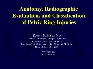
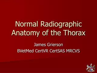
AP ABDOMEN STOMACH COLON SM. BOWEL Normal abdominal gas pattern with air in the stomach and scattered non-distended loops of large and small bowel.
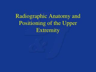
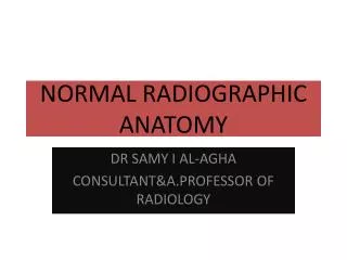
Barium swallow, esophagus. Oblique view • The normal impressions made by : • (A) aortic arch, • (B) left mainstem bronchus, and • (LA) left atrium on the esophagus.
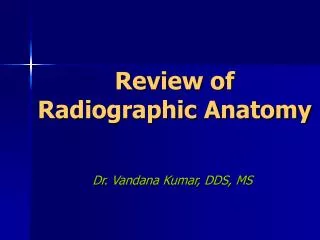
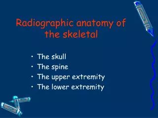
Barium Meal FUNDUS NORMAL GASTRIC ANATOMY DUODENUM ANTRUM BODY JEJUNUM C-LOOP
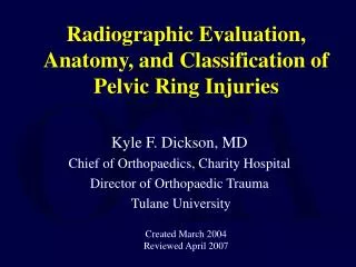
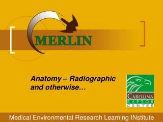
SPLENIC FLEXURE HEPATIC FLEXURE Barium Enema TRANSVERSE COLON DESENDINGCOLON ASCENDING COLON NORMAL COLON TERMINAL ILEUM CECUM