Download
1 / 19
380 likes | 937 Vues
Anna Dubiel. Principles of fluorescent probes. Warsaw , 17.05.2011. Plan of presentation. -> introduction in fluorescence microscopy -> basics of fluorescence -> properties of fluorescent probes -> cell probing: > DNA > membranes > tubulin > mitochondria > indicators for Ca 2+

E N D
Anna Dubiel Principles of fluorescentprobes Warsaw, 17.05.2011
Plan of presentation -> introduction in fluorescence microscopy -> basics of fluorescence -> properties of fluorescent probes -> cell probing: > DNA > membranes > tubulin > mitochondria > indicators for Ca2+ > pH -> summary
Disadvantages of optical microscopy • Usual microscopes working at visible spectral range have magnification coefficient up to 1 500x • Microscopes for ultra-violet have magnification coefficient up to 3500x • Majority of cell components are transparent and colourless • Dyes used in traditional microscopy often are not selective and paint whole cell
Advantages of fluorescent microscopy • Higher sensitivity • Using visible or near IR spectral range • Dyes which are specific for subcellular components, proteins or ions • Observation of cell division • 3D
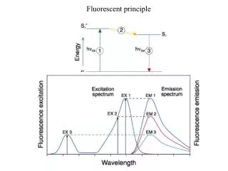
Basics of fluorescence Jabłoński diagram Aleksander Jabłoński 1898 – 1980 Profesor of Nicolaus СoperniсusUniversity in Torun IC + VR IC + VR
Basics of fluorescence Jabłoński diagram IC + VR Absorption: IC + VR Fluorescent quantum yield: Brightness: Stokes shift:
Dyes: • High absorption • High fluorescent quantum yield • Water solubility • Affinity to a particular part of the cell • Chemical and photostability • Stability in cell conditions • Low cytotoxicity
Dyes for imaging: Rhodaminedyes Fluoresceines fluoresceine λabs=489nm λem=534nm ϕf=0,73 ε= 92 300 M-1cm-1 rhodamine B λabs=542nm λem=579nm ϕf=0,50 ε= 106 000 M-1cm-1 J. Mater. Chem., 2009, 19, 2018–2025
Dyes for imaging : Cyanines Coumarins coumarin 440 λabs=354nm λem=434nm ϕf=0,73 ε= 23 500M-1cm-1 Cy3 λabs=546nm λem=571nm ϕf=0,05 ε= 271 000M-1cm-1 Angew. Chem. Int. Ed. 2009, 48, 299 –303 Chem. Phys. Lett., 1978, 54, 159-163 Chem. Med. Chem. 2010, 5, 103 – 117
Dyes for imaging : BODIPY 4,4-difluoro-4-bora-3a,4a-diaza-s-indacene λabs=499nm λem=535nm ϕf=0,93 ε> 80 000 M-1cm-1 J. Org. Chem. 2009, 74, 5719–5722
DNA detection DAPI Λabs=358nm λem=461nm ϕf=20% Cy5-dUTP λabs=649nm λem=670nm ϕf=28% The MolecularProbesHandbook; A Guide to fluorescentProbes and Labelingtechnologies; Eleventh Edition Ewa M. Goldys, FluorescenceAplicationsinBiotechnology and the Life Science; Wiley-Blackwell; 2009
Probes for membranes ß-BODIPY FL C5-HPA λabs=504nm λem=511nm ε= 79 000 M-1cm-1 Fluorescein DHPE λabs=496nm λem=519nm ε= 88 000 M-1cm-1
Probes for tubulin Oregon Green 488 Taxol λabs=494nm λem=522nm ε= 80 000 M-1cm-1 BODIPY FL vinblastine λabs=503nm λem=510nm ε= 83 000 M-1cm-1
Probes for mitochondia MitoTracker Green FM λabs=490nm λem=516nm ε= 119 000 M-1cm-1 MitoTracker Red CMXRos λabs=578nm λem=599nm ε= 116 000 M-1cm-1
Indicators for Ca2+ Fura Red λabs=473, 436nm λem=670, 655nm ε= 29 000, 41 000 M-1cm-1 Calcium Orange λabs=549nm λem=575nm ε= 80 000 M-1cm-1
pHindicators 5-(and-6)-carboxy SNARF-1 λabs=548, 576nm λem=587, 635nm ε= 27 000, 48 000 M-1cm-1
Summary • Fluorescence microscopy: • -> An essential tool in biology and the biomedical sciences • -> Based on fluorescence fenomena • -> Use fluorescent probes • Aplication of fluorescence microscopy: • Detection and determination of the proteins localization in cell and tissue • Examination changes of ions concetration • Diagnostic of diseases
Thank you for your attention