10
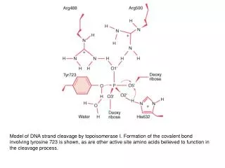
Model of DNA strand cleavage by topoisomerase I. Formation of the covalent bond involving tyrosine 723 is shown, as are other active site amino acids believed to function in the cleavage process.

10
E N D
Presentation Transcript
Model of DNA strand cleavage by topoisomerase I. Formation of the covalent bond involving tyrosine 723 is shown, as are other active site amino acids believed to function in the cleavage process.
A model for topoisomerase II action. As indicated, ATP binding to the two ATPase domains causes them to dimerize and drives the reactions shown. Because a single cycle of this reaction can occur in the presence of a non-hydrolyzable ATP analog, ATP hydrolysis is thought to be needed only to reset the enzyme for each new reaction cycle. This model is based on structural and mechanistic studies of the enzyme
Structure of a Topoisomerase. The structure of a complex between a fragment of human topoisomerase I and DNA.
Structure of Topoisomerase II. A composite structure of topoisomerase II formed from the amino-terminal ATP-binding domain of E. coli topoisomerase II (green) and the carboxyl-terminal fragment from yeast topoisomerase II (yellow). Both units form dimeric structures as shown.
The mode of action of Type I and Type II DNA topoisomerases. (A) A Type I topoisomerase makes a nick in one strand of a DNA molecule, passes the intact strand through the nick, and reseals the gap. (B) A Type II topoisomerase makes a double-stranded break in the double helix, creating a gate through which a second segment of the helix is passed.
Topoisomerase I Mechanism. On binding to DNA, topoisomerase I cleaves one strand of the DNA through a tyrosine (Y) residue attacking a phosphate. When the strand has been cleaved, it rotates in a controlled manner around the other strand. The reaction is completed by religation of the cleaved strand. This process results in partial or complete relaxation of a supercoiled plasmid.
The DNA-helix-passing reaction catalyzed by DNA topoisomerase II. Identical reactions are used to untangle DNA inside the cell. Unlike type I topoisomerases, type II enzymes use ATP hydrolysis and some of the bacterial versions can introduce superhelical tension into DNA. Type II topoisomerases are largely confined to proliferating cells in eucaryotes; partly for that reason, they have been popular targets for anticancer drugs.
Model of topoisomerase 2 catalysis. The DNA duplex that undergoes cleavage is referred to as the G-segment (for “gate”) and the other DNA duplex is referred to as the T-segment (for “transported”). Binding of the G-segment (step 1) results in a conformational change (step 2) in which the active site tyrosines (shown as purple circles) are brought into position for cleavage of the G-segment. After binding of the T-segment and ATP, a “clamp” is formed around the T-segment (step 3), which is then transported though the gap in the G-segment (step 4). Subsequently, the G-strand is religated and the Tsegment is released (step 5). After ATP hydrolysis, the “clamp” is opened and the cycle can repeat
Linking Number. The relations between the linking number (Lk), twisting number (Tw), and writhing number (Wr) of a circular DNA molecule revealed schematically