Download
1 / 1
10 likes | 192 Vues
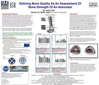
Defining Bone Quality As An Assessment Of Bone Strength Of An Astronaut. By: Ashley Titan Mentors: Dr. Stefan Judex, Dr. Steven Tommasini. BACKGROUND INFORMATION

E N D
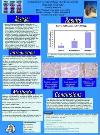
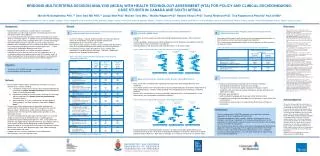
Defining Bone Quality As An Assessment Of Bone Strength Of An Astronaut By: Ashley TitanMentors: Dr. Stefan Judex, Dr. Steven Tommasini BACKGROUND INFORMATION The skeleton has evolved to allow efficient locomotion and support the body [1]. Bones are made up of two kinds of tissue: Trabecular bone is highly porous with low density and high surface area [2]. Cortical Bone is the very dense outer shell [2]. The strength of bone is based its quantity, shape, thickness, and composition. It is important to note that it is essential to understand both bone quantity and quality when defining bone strength. Due to gravitational differences the human skeleton does not need the same amount of strength in outer space as on Earth. In previous studies it has been shown that both cortical and trabecular bone mineral loss results from long-duration spaceflight. Some bone specimens lost up to twice the amount of mineral content compared to there earthly counterparts [3]. Existing bone strength diagnostic methods are unreliable. Susceptibility to bone fracture is currently measured by Bone Mineral Density (BMD). This parameter accounts for < 40% of variation in trabecular bone strength [4]. The goal of this study is to determine more accurate ways to analyze and define bone strength. It is proposed that the strength and quality of the bone’s matrix will be significantly influenced by both the chemical and structural composition. CONCLUSIONS As expected there were a myriad of differences between OVX and control groups. There were also similarities between the treatment and control groups. A decrease in Ct.Area was seen when comparing the controls to the OVX group, however, an increase in Ct.Area was noted in the OVX group that received treatment especially with 100ug ALN (Graph 1). When looking at BV/TV and Ct.Th it was apparent that the OVX bone was thinner than the control bone, while the bones of OVX group that received treatment were similar in thickness to the control bone (Graphs 2&4). Lastly, the TMD parameter was greater for the OVX group than the control, but with treatment the OVX values for TMD were reduced towards the control level (Graph 3). These analyses are made visible as seen in Figure1 where images of the distal metaphysis from both the OVX and control groups are placed next to one and other. It should be noted that none of these trends were shown to be statistically significant. However, with an increase in sample size and the amount of time periods this should be subject to change. Bigger differences are expected between groups when analyzing trabecular bone (Figure 1). Treatments used in this study may in the long-term be used in medicine to increase bone strength of patients who develop weak bones such as individuals with osteoporosis and astronauts who need to travel for extended periods of time. Regions of Interest: Results: Ct.Area BV/TV • METHODS AND MATERIALS • Rat femora were divided into experimental groups based on age (2 to 8 • month olds), treatment (OVX -Ovariectomy, ALN-Alendronate, PTH- • Parathyroid Hormone, and control). • The left femur was used to study the microscopic level for correlations • between chemical and structural properties: • - FTIR - chemical properties • - Nanoindentation - microscopic mechanical properties • - MicroCT - structural properties • The right femur was used to study mechanical behavior at the • macroscopic level. • Trends were then evaluated in a variety of categories including • volumes, perimeters, areas, bone volume fraction, moment of inertia, • bone mineral, porosity, and cortical thickness. • Statistical analyses were performed on Graphpad Prism 5.0 and • Microsoft Excel. Right Femur Left Femur MicroCT Mechanical Testing Anterior MicroCT FTIR NanoCT Nanoindentation Anterior Lateral Medial Lateral Medial Graph 1: There is a noticeable difference for Ct.Area between OVX and control groups as well as similarities between the control and most treatment groups. Graph 2: The OVX showed bone loss compared to controls, whereas most treatment groups have similar bone mass to controls. Posterior Posterior TMD Ct.Th OVX Distal Metaphysis Control Distal Metaphysis Figure 1: There are apparent differences in the quality and quantity of the bone between the OVX and control bones. Future Research Further work needs to be done to understand bone quality and treatments in order to improve bone strength. This would be completed through FTIR, Nano-indentation, NanoCT testing. Increasing sample size and the amount of time periods would likely cause significant differences between the OVX, control and treatment groups. Analysis of the trabecular MicroCT data will also be completed so demonstrate significant differences between groups. Graph 3: Control TMD was greater than the OVX group. Additionally, the treatment groups have TMD values similar to control group. Graph 4: Control Ct.Th was greater than the OVX group. Additionally, the treatment groups have Ct.Th values similar to control group. Sponsors: National Aeronautics and Space Administration (NASA) NASA Goddard Space Flight Center (GSFC) NASA Goddard Institute for Space Studies (GISS) NASA New York City Research Initiative (NYCRI) SUNY Stony Brook University Contributors: Dr. Stefan Judex Dr. Steven Tommasini Svetlana Lublinsky Ashley Titan REFERENCES 1. Leppänen, O., H. Sievänen, and T. Järvinen. “Biomechanical testing in experimental bone interventions – May the power be with you.” Journal of Biomechanics. (2008). 2. "Biomechanics." BME. 2007. University of Michigan. 20 Oct. (2007). <http://www.engin.umich.edu/class/bme456/bonestructure/bonestructure.htm>. 3. Lang, T., A. LeBlanc, H. Evans, Y. Lu, H. Genant, and A. Yu. “Cortical and trabecular bone mineral loss from the spine and hip in long-duration spaceflight.” Journal of Bone and Mineral Research. (2008). 4. Mc Donnell, P., M. Liebschner, W. Tawackoli, P. Hugh. “Vibrational testing of trabecular bone architectures using rapid prototype models.” Medical Engineering and Physics. (2008).