Download
1 / 61
730 likes | 1.15k Vues
Blood Biochemistry. Composition of Blood. Blood is the body’s only fluid tissue It is composed of liquid plasma and formed elements Formed elements include: Erythrocytes, or red blood cells (RBCs) Leukocytes, or white blood cells (WBCs) Platelets

E N D
Composition of Blood • Blood is the body’s only fluid tissue • It is composed of liquid plasma and formed elements • Formed elements include: • Erythrocytes, or red blood cells (RBCs) • Leukocytes, or white blood cells (WBCs) • Platelets • Hematocrit – the percentage of RBCs out of the total blood volume
Plasma(55% of whole blood) Formed elements Erythrocytes(45% of whole blood) Components of Whole Blood Buffy coat:leukocyctes and platelets(<1% of whole blood) Withdraw blood and place in tube Centrifuge 1 2
Plasma • The blood fraction obtained after removal of the cellular components • About 77%-81% in the total blood values • Hydrometer is 1.050-1.060, pH is 7.35-7.45, osmotic pressure is 770kPa (37°C) in the normal human • relevant to coagulation factors, immunoglobulins and complements
Serum • The blood fraction after separation of the protein fibrinogen from plasma • Generally obtained by allowing the blood to clot • In this process, fibrinogen is converted to an insoluble protein, fibrin, which is easily removed • Serum does contain some degradation products of clotting factors
Plasma composition Clotting factors Liquid: water protein plasma Nonprotein nitrogen (NPN) Low-molecular-weight organic substances such as Serum solids Serum solids glucose, lipids, vitamins, hormones and so on Na+, K+, Ca2+, Mg2+ electrolytes CI-, HCO32-, HPO42- Gases:O2, CO2
Non-Protein Notrogen (NPN) • Non-protein nitrogenous compounds urea, uric acid, creatinine, creatine, nucleotides, amino acids, bilirubin, polypeptides, glutathione and many others • The Concentration of NPN 14.28~24.99 mmol/L, 50% of NPN is blood urea nitrogen (BUN) • Source of NPN derived from the metabolism of nucleic acid and proteins • Excretion of NPN transported to the kidneys fro excretion from the urin • Significance act as an index of renal function
Male versus female • Hematocrit (% volume that is red cells) • 40-50% in males • 35-45% in females
Function of Blood • Blood as a transport system transport nutrients and oxygen to the cells and carries away cellular waster products • Blood as a regulative system maintaining normal acid-base balance in the body; Regulating the water balance and body temperature • Blood as a defense system white blood cells and the circulating antibodies • Coagulation and fibrinolysis
Section 1 Plasma Proteins
Plasma Proteins • More than 200 • Most abundant • Albumin - 4-5 g/100 mL • g-glubulins - ~1 g/100 mL • fibrinogen - 0.2-0.4g/100 mL • Original classification by zone electrophoresis at pH 8.6 • Separation by pI with several molecular weight species within each group
Zone Electrophoresis of Plasma Proteins + - globulins albumin g b a1 a2 pI 6.0 5.6 5.1 4.7
Protein Separation • Size Exclusion Chromatography (SEC) • Porous matrix (sephadex)
Affinity chromatography • molecule attached to a column that specifically binds the protein of interest • Coenzyme / enzyme • Antigen / Antibody
SDS-PAGE (polyacrylamide gel electrophoresis) • Separates by size • Proteins are complexed with SDS to give the same charge density
Two Dimensional Electrophoresis Decreasing Mr Decreasing pI
Characteristics of Plasma Proteins • Most plasma proteins are synthesized in the liver, however, certain proteins are synthesized in other sides • Generally synthesized on membrane –bound polyribosomes • With the exception of albumin, almost all plasma proteins are glycoproteins • Many plasma proteins exhibit polymorphism • Each plasma protein has a characteristic half-life in the circulation • The levels of certain proteins in plasma increase during acute inflammatory states or secondary to certain types of tissue damage
Functions of Plasma Proteins (1) Functional enzymes of the plasma • Have catalysis in the plasma, such as thrombin, lipoprotein lipase, LCAT etc
(2) non-functional enzymes of the plasma • Maintenance of: • Colloid osmotic pressure (COP) (p) • pH • electrolyte balance • COP relates to blood volume DP = p Protein sol’n Water
Transport of ions, fatty acids, steroids, hormones etc. • Albumin (fatty acids), ceruloplasmin (Cu2+), transferrin (Fe), lipoproteins (LDL, HDL) • Nutritional source of amino acids for tissues • Hemostasis (coagulation proteins) • Prevention of thrombosis (anticoagulant proteins) • Defense against infection (antibodies, complement proteins)
80 Å 30 Å Albumin • MW 66 000 • Single chain, 580 amino acids, sequence is known • Dimensions - Heart shaped molecule • 50% a helix [He and Carter, Nature, 358 209 (1992)] • Modeled as:
Synthesis • Mainly liver cells then exported • Assembly time on ribosome ~ 1-2 min • t0.5 in circulation - 19 days • 14 g lost per day • 0.4 mg synthesized per hour per g of liver • Need liver of approximately 1.5 kg in weight to maintain
Functions • Maintaining colloid osmotic pressure of blood (80% due to albumin) • Colloid osmotic pressure is generated by plasma proteins • The most abundant of the plasma proteins • The lowest molecular weight of the major protein molecules in the plasma • High negative charge • Regulates water distribution • Transportation • Albumin can act as a carrier molecule for bilirubin, fatty acids, trace elements and many drugs
Section 3 Metabolism of the Blood Cells
Cellular Elements of Blood • Red cells • 40 - 50% of blood volume • 5 x 10⁶ cells /mL • Composed of a membrane surrounding a solution of hemoglobin • non-nucleated, no intracellular organelles • no proliferation • cell membrane in excess so that deformation does not rupture • Shape • Biconcave disc • 8 mm in diameter, 2.7 mm thick, volume ~ 90 mm3, area ~ 160 mm2
Why this shape? • Area to volume ratio is high • Facilitates diffusion of O2 and CO2 • minimal distance of contents from surface • Originates in bone marrow (hematopoiesis) • Molecular explanation based on the properties of the proteins in the cell membrane is found in Elgsaeter et al. Science, 234, 1217 (1986)
Production of Erythrocytes • Hematopoiesis – blood cell formation • Hematopoiesis occurs in the red bone marrow of the: • Axial skeleton and girdles • Epiphyses of the humerus and femur • Hemocytoblasts give rise to all formed elements
Production of Erythrocytes: Erythropoiesis • A hemocytoblast is transformed into a committed cell called the proerythroblast • Proerythroblasts develop into early erythroblasts • The developmental pathway consists of three phases • Phase 1 – ribosome synthesis in early erythroblasts • Phase 2 – hemoglobin accumulation in late erythroblasts and normoblasts • Phase 3 – ejection of the nucleus from normoblasts and formation of reticulocytes • Reticulocytes then become mature erythrocytes
Production of Erythrocytes: Erythropoiesis Figure 17.5
The major function of the red cells • Delivering oxygen to the tissues, helping in the disposal of carbon dioxide and protons formed by tissue metabolism
Normal red cell breakdown haemoglobin haem globin iron protoporphyrin Amino acids Bilirubin (free) CO Expired air transferrin Liver conjugation erythroblast Bilirubin glucuronides Urobilin(ogen) Stercobilin(ogen) Urine faeces
HSCoA + CO2 ALA synthase ( Pyridoxal phosphate) Hemoglobin synthesis • Heme synthesis starts with the condensation of glycine and succinyl coenzyme A under the action of a rate limiting enzyme δ-aminolevulinic acid (ALA) synthase. • δ -ALA will be formed. • Pyridoxal phosphate (vit. B6) is a coenzyme for this reaction. + This step takes place in the mitochondria
ALA dehydratase 2H2O A series of biochemical reactions will follow.Two molecules of δ-ALA condense to form a pyrrole called porphobilinogen (PBG) This step occurs in the cytoplasm
Four PBG condense to form a tetrapyrrole uroporphyrinogen III. • UPG III is then converted to coproporphyrinogen. Linear tetrapyrrole Deaminase Four PBG UPG III isomeiase UPG III decarboxylase coproporphyrinogenⅢ uroporphyrinogen III This step occurs in the cytoplasm
Haemoglobin synthesis • CPG then changes to protoporphyrin which ultimately combines with iron in the ferrous state (Fe2+) to form haem. • Iron is brought to the developing red cells by a carrier protein ( transferrin) which attaches to special binding sites on the surface of these cells. • Transferrin releases iron and returns back to circulation.
Haemoglobin synthesis • Each molecule of haem combines with a globin chain. • A tetramer of four globin chains each with its own haem group in a pocket is formed to make up a haemoglobin molecule.
Haemoglobin structure • Haem consists of a protoporphyrin ring with an iron atom at its centre. • The protoporphyrin ring consists of four pyrrole groups which are united by methane bridges (=C-). • The hydrogen atoms in the pyrrole groups are replaced by four methylene (CH3-), two vinyl (-C=CH2) and two propionic acid (-CH2-CH2-COOH) groups.
Metabolic Characteristics of Mature Erythrocytes • Can not carry out synthesis of nucleic acid and proteins • Can not obtain energy by oxidative phosphorylation of the mitochondria • ATP is synthesized from glycolysis and is important in process that help the red blood cell maintain its biconcave shape and also in the regulation of the transport of ions and of water in and out of the cell • The principal modes of glucose metabolism are anaerobic glycolysis and the pentose-phosphate pathway
Glycolysis • Obtain energy by glycolysis of glucose Utilize 2ATP moleculars, produces 4ATP moleculars with a net gain of 2ATP • The function of ATP Tomaintain the correct ion balance, brought about by the pumping out of sodium in exchange for potassium To maintain the correct conformation of the cell To protect against the formation of methaemoglobin To synthesize NAD+ and glutathione
The pathway of 2,3-bisphosphoglycerate (2,3-BPG) • Formation of 2,3-BPG Glucose Diphosphoglyceromutase 1, 3-BPG Phosphoglycerate kinase 2, 3-BPG Glycerate 3-phosphate Diphosphoglycerate phosphatase Lactate
The role of 2,3-BPG • Plays an important role in the binding of oxygen to hemoglobin in erythrocytes • Combine with hemoglobin, causing a decrease affinity of hemoglobin for oxygen pO2 2,3-DPG (lungs) Oxyhemoglobin Hemoglobin pO2 2,3-DPG (tissues) (HbO2) (Hb)
NADP+ 2GSH Glutathione reductase NADP++H+ GSSG Pentose phosphate pathway The role of the pentose phosphate pathway • Produce the NADH which is essential for the regeneration of reduced glutathione from oxidized glutathione
NADP+ 2GSH H2O2 Glutathione reductase Glutathione peroxidase 2H2O NADP++H+ GSSG The role of glutathione are as follows The role in the destruction of hydrogen peroxide (H2O2) in erythrocytes
Reduction of methemoglobin • Methemoglobin does not combine with molecular oxygen and does not have the function of transporting oxygen • Normally, methemoglobin is reduced to the ferrous state by the NADH-dependent methrmoglobin reductase Methrmoglobin reductase MHb (Fe3+) Hb (Fe2+) NAD H2O ½ O2 NADH+H+
Genetic abnormality-deficiency of glucose-6-phosphate dehydrogenase • glucose-6-phosphate dehydrogenase is the first enzyme of the pentose phosphate pathway • A deficiency of this enzyme will lead to failure of restoring GSSG to GSH in the erythrocytes, a step essential for the removel of H2O2 • Cell damage is likely to result from oxidation of the membranes by the H2O2, leading to hemolytic anemia
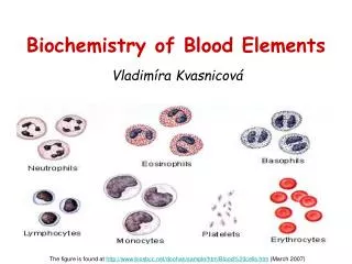
White Blood Cells (Leukocytes) • Total count - approximately 7000/mL • Various types • Neutrophils 62% • Eosinophils 2.3% • Basophils 0.4% • Monocytes 5.3% • Lymphocytes 30% • Plasma cells (mainly in the lymph) • Monocytes in tissue become macrophages granulocytes
Function • Defense against foreign invaders • bacteria • viruses • foreign materials (including biomaterials) • Phagocytosis • Neutrophils, macrophages • Move to foreign particle by chemtaxis • Chemicals induce migration • Toxins, products of inflamed tissues, complement reaction products, blot clotting products • Response is extremely rapid (approx 1 h)
Lymphocytes • B cells - responsible for humoral immunity • T cells - responsible for cell mediated immunity • B cells responsible for production of antibodies • Receptor matches antigen • Cells multiply • Antibodies • Abs are just immunoglobulins discussed earlier