Download
1 / 57
1.73k likes | 4.77k Vues
Biochemistry of Blood. Bruno Sopko. Content. Introduction Blood Plasma Metabolism of E rythrocytes Metabolism of White Cells Phagocytic cells Basophils and mast cells Lymphocytes Biochemistry of Platelets / Blood Coagulation Literature. Introduction. Functions

E N D
Biochemistry ofBlood Bruno Sopko
Content • Introduction • Blood Plasma • Metabolism of Erythrocytes • Metabolism of WhiteCells • Phagocyticcells • Basophilsand mast cells • Lymphocytes • Biochemistryof Platelets/Blood Coagulation • Literature
Introduction • Functions • Blood Composition
Introduction – Functions I. • Respiratory • CO2 transport from tissues to lungs • O2 transport from lungs to tissues • Nutrition • Transports nutrients from digestion system to tissues • Excretory • Transports waste from tissues to kidneys (urea, uric acid, water, salts etc.)
Introduction – Functions II. • Regulatory • Watercontent in thetissues • Distributionoftheregulatorycompounds(hormons etc.) • Body Temperature • Water has high heat capacity (heat accumulation) • Heat spreading from one source (cooling, warming) • Protective • Antibodies, antitoxins, white blood cells
Introduction – Blood Composition • 8% of the body weight (5–6 L) • Suspension of cells in carrier fluid • 45% cells 55% plasma • Plasma • Red Cells • White cells • Platelets
Plasma I. - Composition • Water (90%) • Proteins (7%) • Most of them synthesized in liver • Mostly polymorfousglycoproteins • Each has specific half-time in circulation (albumin 20 days, haptoglobin 5 days • Concentration of some of them changes during inflammation (acute-phase proteins) • Inorganic (1%, Na+, K+, Mg2+, Ca2+, PO43-, Cl-...) • Organic (2%, urea, fats, cholesterol, glucose, aminoacids …)
Erythrocytes 5.2 ×10⁶ (men); 4.6 ×10⁶ women cells/ml
Hemoglobin autooxidation • O2 binds Fe2+ - an intermediate structure - an electron is delocalized between the iron ion and the O2 • the side effect - every so often a molecule of oxyhaemoglobin undergoes decomposition and release superoxide Hem - Fe2+- O2 Hem - Fe3+ - O2•- • 3% of the haemoglobin undergoes oxidation every day • Methemoglobin (Fe3+) is unable to bind O2 (methaemoglobinreductase)
The pentose phosphate pathway in erythrocytes • Generates NADPH - reduction of glutathione (eliminates H2O2 formed in erythrocytes) Clinical apect: • Glucose-6-phosphate dehydrogenase deficiency • Causes hemolytic anemia (decreased production of NADPH - reduced protection against oxidative stress - haemoglobin oxidation and Heinz bodies formation, membrane lipid peroxidation and hemolysis) • Hemolytic crises are evocated by drugs (primaquine, sulphonamide antibiotics) and foods (broad beans) • The most common enzyme deficiency disease in the world (100 million people)
Haemoglobinopathies Haemoglobinopathy • abnormalstructureofthehaemoglobin (mutation) • largenumberofhaemoglobinmutations, a fraction has deleteriouseffects • sickling, change in O2affinity, heme lossordissociationoftetramer • haemoglobin M and S, andthalassemias Haemoglobin M • replacementofthe histidine (E8 or F7) in αorβ-chain by thetyrosine • the iron in the heme groupis in the Fe3+state(methaemoglobin) stabilized by thetyrosine • methaemoglobincan not bind oxygen Thalassemias • geneticdefects- αorβ-chains are not produced(αorβ-thalassemia) HaemoglobinS (sickle-cell) • Causes a sickle-cell anemia • Erythrocytesadoptanelongatedsickleshapedue to theaggregationofthehaemoglobin S
Glycosylatedhaemoglobin (HbA1) • formed by hemoglobin's exposure to high plasma levels of glucose • non-enzymatic glycolysation (glycation)- sugar bonding to a protein • normal level HbA1- 5%; a buildup of HbA1- increased glucose concentration • the HbA1 level is proportional to average blood glucose concentration over previous weeks; in individuals with poorly controlled diabetes, increases in the quantities of these glycatedhemoglobins are noted (patients monitoring) Sugar CHO + NH2 CH2 Protein Sugar CH N CH2 Protein Sugar CH2 NH CH2 Protein Schiff base Amadorireaction Glycosylated protein
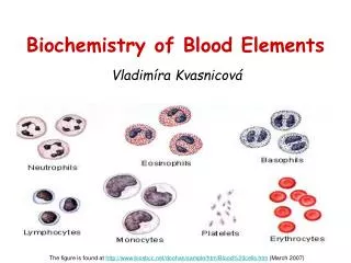
White Blood cells • Phagocyticcells • Basophilsand mast cells • Lymphocytes
Phagocyticcells • Introduction • Granulocytes • Neutrophils– most abundant • Eosinophils • Basophils • Monocytes • Macrophages – rise by differentiationofmonocytes in tissues
Degradation of the ingested particle 1) Activation of NADPH oxidase 2) Production of NO by nitric oxide synthase 3) Fusion of phagosome with lysosomes of the phagocytic cell that contain bactericidal substances and hydrolytic enzymes (often with acidic pHopt)
NADPH-oxidase • Protein complexofneutrophils, eosinophils, monocytes, macrophages NADPH + 2 O2→ NADP+ + H+ + 2 O2•- 2 O2•- + 2 H+ → O2 + H2O2 • H2O2 candamagebacteriadirectlyorafterconversion to OH• : H2O2 + M+ → OH• + OH- + M2+(M; metal) superoxide anion
NADPH-oxidase • Activation: by association of the components localized in cytosol with cytochrome b558 in the membrane; electrons from cytosolic NADPH are – via FAD and cytochrome – transferred to oxygen
NADPH-oxidase plasma membrane fusion with lysosomes phagosome
Myeloperoxidase • Present in granules of neutrophils and monocytes, but not macrophages! • Significant portion of H2O2 (produced by dismutation of O2•- generated by NADPH oxidase) is used by myeloperoxidase to oxidize Cl- to HClO • HClO is highly reactive, able to oxidize biomolecules; it also provides toxic chlorine gas: HClO + H+ + Cl-→ Cl2 + H2O • HClO also reacts with O2•- yielding OH•: HClO + O2•- → O2 + OH• + Cl-
Chronic granulomatous disease • Caused by a deficiency of one of the NADPH oxidase subunits • Superoxide and the other reactive oxygen species are not produced • Severe infections that are very hard to treat – e.g.: • Burkholdaria cepacea causes pneumonia • Aspergillus causes intractable pneumonia, septicaemia; can lead to death • Treatment: antibiotics, antifungal agents
Nitric oxide production • Mainly by inducible nitric oxide synthase (iNOS) of macrophages which is induced by cytokines (INF-γ, TNF)or bacterial lipopolysaccharide: • NO• can kill bacteria directly (e.g. by inhibition of the respiratory chain) or indirectly: by reaction with O2•-, generating peroxynitrite ONOO- which attacks Fe-S proteins and essential –SH groups, inactivates enzymes… Arg citrulline
NADPH oxidaseandNO • NADPH oxidase is effective mainly in degradation of extracellular pathogens (Salmonella, Staphylococcus, Streptococcuspyogenes)…neutrophils X • NO serves mainly to kill the intracellular parasites (Listeria, Brucella, Candida albicans)…macrophages
Granulocytes - introduction • All of them are phagocytes • Contain several types of granules • Primary granules – participating in phagocytosis • Secondary granules – releasing cytotoxic and immune response mediators (defensins, cathepsins etc.) • After phagocytosis is accomplished, the respiratory burst occurs
Neutrophils • 40 – 65 % of white blood cells • Must be activated • React mostly with opsonised cells • Mediate other immune response (eicosanoids, cytokines)
Neutrophils Maintargets are bacteria • Myeloperoxidase • lysozyme – cleaves glycosidic bonds in peptidoglycan of the bacterial (primarily G+) cell walls • defensins – cationic peptides (Arg) with Mr of 3,5-6 kDa; interact with anionic lipids of bacterial membrane and make pores in it; can also inhibit synthesis of DNA and proteins • hydrolases, e.g. elastase – serine protease: can damage bacteria and cleave virulence factors, but also cause harm to host tissues (cleaves the proteins of extracellular matrix, too)
Eosinophils Maintargets are eucaryoticParasites • ROS production • Contain eosinophilperoxidase • similar to myeloperoxidase, but prefers Br-as a substrate (instead of Cl-), thus generating HBrO(instead of HClO) • proteases • Granules contain Major Basic Protein, cytotoxic to parasites • Release of histamine
Basophils • Mainly immune response mediator (histamine and serotonin) • Contain IgE receptors, once activated, degranulate • responsible for allergic symptoms • Activate synthesisof eicosanoids; leukotrienes are potent bronchoconstrictors, stimulatechemotaxisand leukocyte activation
Histamine • Produced by histidine decarboxylation: • Causes vasodilationand bronchoconstriction helps to eliminate parasites (cough, peristalsis, enhanced production of mucus)
Atopy • IgE recognizing allergens (from pollen, food…) are produced and bind toIgE receptors of basophils (mast cells). Next exposure tothe allergen can lead to release of histamine and heparin and synthesisofeicosanoids • Local symptoms occur: allergic rhinitis, asthma, conjunctivitis • If the allergen enters bloodstream, it can cause a massive degranulation of basophils (mast cells) increase in vascular permeability, decrease in blood pressure pulmonary oedema, ischemia… anaphylacticshock • Treatment: antihistamines – block histamine receptors
Lyphocytes • T cells • B cells • NK cells
Fixed leukocytes • Histiocytes • Dendritic Cells • Mast Cells • Microglia • Kupffer Cells (liver)
Blood coagulation - platelets • Non nucleated • Granulated • electron-dense granules, which contain calcium, adenosine diphosphate (ADP), adenosine triphosphate (ATP), and serotonin • granule, which contains a heparin antagonist (heparin interferes with blood clotting; see biochemical comments), platelet-derived growth factor, -thromboglobulin, fibrinogen, von Willebrand factor (vWF), and other clotting factors • the lysosomal granule, which contains hydrolytic enzymes
Regulations • Many functions of leukocytes are regulated by monomeric GTP-binding proteins, e.g. Rac, Rho: • activation of NADHP oxidase • chemotaxis • phagocytosis • fusion of phagosome with granules • Rho and Rac are able to modulate the assembly of actin filaments, which plays a role in the processes listed above
Platelets • Form blood clots, act as vasoconstrictors • Participate in defence against infections, e.g.: they suppress the growth of Plasmodium falciparum (infectious agent that causes malaria) • Generate O2•- and H2O2 that may synergize with pro-aggregatory stimuli • Contain thromboxan A synthase that catalyzes conversion of prosta-glandin H2 to thromboxan A2: TXA2 – promotes platelet aggre- gation and vasoconstriction
Platelets • Platelet-Activating Factor (PAF) • Platelet-Derived Growth Factor (PDGF)
Platelet-Activating Factor phospholipid • Mainly juxtacrine and paracrine signalling via GPCR • Promotes platelet aggregation • Induces activation of leukocytes, adhesion, chemotaxis, cytokine production, causes vasodilation and bronchoconstriction • Mediates interplay between thrombotic and inflammatory cascades • BUT: it is also suspected of contributing to allergy, anaphylactic shock… • It is produced also by endothelial cells, monocytes, granulocytes…