Craniofacial and Pharyngeal Arch Development
721 likes | 2.34k Vues
Craniofacial and Pharyngeal Arch Development. Matthew Velkey matt.velkey@duke.edu 454D Davison. Two general phases of pharyngeal apparatus development. Wk 3.5. Wk 4.5. Wk 5.5. Wk 6.5. Disruptions to either phase can result in a broad spectrum of defects.

Craniofacial and Pharyngeal Arch Development
E N D
Presentation Transcript
Craniofacial and Pharyngeal Arch Development Matthew Velkey matt.velkey@duke.edu 454D Davison
Two general phases of pharyngeal apparatus development Wk 3.5 Wk 4.5 Wk 5.5 Wk 6.5 Disruptions to either phase can result in a broad spectrum of defects.
Early pharyngeal apparatus development • 5 pairs arise in cranial to caudal direction • Arch 1 = Mandible • Arch 2-4, 6 = Neck • No Arch 5 in mammals
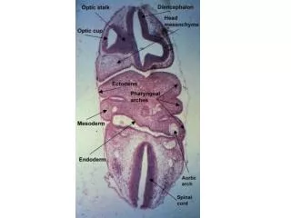
Organization & components of pharyngeal arches plane of section
Neural Crest Mesoderm Ectoderm In vivo marking of pharyngeal arch components
Coincident development of related structures within the pharyngeal apparatus • Significance: • Understanding the origins of developmentally-related structures • A genetic lesion affecting a particular pharyngeal arch will frequently affect multiple developmentally related structures
1st Arch Defects • Pierre Robin Syndrome: small mandible, cleft palate, middle ear defects, low-set ears • Treacher Collins Syndrome: mutation in Treacle gene, small mandible, middle ear defects, low-set ears, cleft plate, tooth defects
Fates of pharyngeal clefts/grooves • 1st cleft • Develops into the external auditory meatus • Only cleft to yield a normal adult structure • 2nd-4th clefts • Normally overgrown by 2nd pharyngeal arch and epicardial ridge • Ultimately degenerate and do not give rise to definitive mature structures in mammals
Dysgenesis of pharyngeal clefts • Fistula: epithelial tubes that open at both ends • Cyst: pocket because cleft didn’t completely degenerate • Preauricular cysts & fistulas: associated with defects in cleft 1 and/or pouch 1 • Lateral cervical cyst: incomplete overgrowth of clefts 2-4 • Branchial fistulas: defects in clefts 2-4 and/or pouches 2-4
Pharyngeal pouches 1 & 2 have non-endocrine fates • 1st pouch: • Proximal: auditory (eustacian) tube • Distal: tympanic (middle ear) cavity • Extreme distal: tympanic membrane (with mesoderm & ectoderm from 1st pharyngeal cleft) • 2nd pouch: • Supratonsillar fossae of Palatine tonsil • Immune cells come from elsewhere!
Fates of pouches 3 & 4 include endocrine glands • 3rd pouch • Solid superior epithelial mass: inferior parathyroid • Inferior elongation: thymus • 4th pouch • Solid superior epithelial mass: superior parathyroid • Inferior mass: ultimobranchial body (neural crest origin): parafollicular (C) cells of thyroid Each primordium (normally) detaches from site of origin and migrates to site of mature gland
Migration of pouch 3 & 4 primordia to mature organ sites • Thymus: • Ends up inferior to thyroid • Parathyroid III: • Ends up inferior to parathyroid IV in thyroid gland • Parathyroid IV: • Ends up in thyroid, superior to parathyroid III • Postbranchial body (ultimobranchial body): • Migrates into thyroid
Ectopic parathyroid or thymic tissue • Typically benign anatomical anomalies, not accompanied by functional abnormalities
DiGeorge (del22q11) Syndrome (DGS) • 1:4000 live births • Most common human genetic deletion syndrome • Loss of pharyngeal arch structures, particularly 2nd and 3rd arches • Clinical features • Thymic hypoplasia (immunodeficiency) • Hypoparathyroidism (hypocalcemia) • Congenital heart defects • Outflow tract, aortic arch • Velopharyngeal dysfunction +/- cleft palate • Developmental & behavioral problems • Psychiatric disorders
Development of the tongue The tongue forms from ventral swellings in the floor of the pharynx at about the same time as the palate begins to form. Lateral lingual swellings are first visible at about 5 wks, 1 medial swelling. Growth of the tongue mostly by expansion of lateral lingual swellings, some contribution of tuberculum impar Musculature derived from occipital myotomes (QuickTime version) Ant 2/3 of tongue is from 1st arch, posterior 1/3 mostly from 3rd arch
The thyroid gland IS NOT derived from pharyngeal pouches! • Starts as singleplacode (thickening) called the foramen cecum and located in the ventral pharynx just caudal to tongue bud • Elongates caudally as thyroid diverticulum • During migration, tip of thyroid diverticulum expands and bifurcates to form the thyroid gland itself • Developing gland remains transiently attached to site of origin by thyroglossal duct, which then (normally) degenerates
Thyroglossal cysts & sinuses • Origins: failure of the thyroglossal duct to completely pinch off & degenerate • Thyroid arrives at normal position and is usually normal functioning • Can usually be distinguished easily from lateral cervical cysts by their midline position thyroglossal sinus
Congenital hypothyroidism (CH) • Most frequent endocrine disorder in newborns (1:3000 live births) • High TSH levels due to reduced thyroid hormone levels • Clinical significance: effects on CNS development (cretinism) and lungs (low surfactant production) • 85% of CH due to thyroid dysgenesis (TD) (disturbances in thyroid organogenesis) • Includes athyreosis (none), hypoplasia (too small), and ectopia (misplaced)
Cranial neural crest Paraxial/lateral plate mesoderm 3rd arch n. crest 4th arch n. crest The bones of the skull, face, and pharynx are derived from either PARAXIAL mesoderm (orange) or NEURAL CREST (blue & yellow)
Some bones of the skull and face develop via INTRAMEMBRANOUS ossification; some develop via ENDOCHONDRAL ossification: • Base of skull (“chondrocranium,” orange) by endochondral ossification • Superior-anterior skull and face (“membranous cranium,” light blue) by intramembranous ossification • Cartilagenous viscerocranium (green) from neural crest that differentiates into cartilage and then undergoes varying degrees of ossification. paraxial origin neural crest origin
Bones are incomplete At birth: fontanelles Posterior: 3 mos Anterior 1.5 years Will bulge out with increased intracranial pressure (e.g. meningitis) FGF/noggin/BMP signaling determines fusion Disruption of FGF/Noggin/BMP can result in premature fusion (aka craniosynostosis) FGF Nog FGF Nog FGF Nog Nog Nog Nog BMP BMP BMP BMP BMP FGF Nog BMP BMP Nog FGF BMP BMP FGF --| Noggin --| BMP bone growth BMP bone growth [i.e. local FGF allows BMP expression (bone growth) in sutures]
In Apert syndrome the coronal sutures fuse prematurely, and the cranium is abnormally shaped to accommodate the growing brain.
Development of the face (QuickTime version)
Pharyngeal (branchial) arches: earliest primordia of the face
Frontonasal prominence (cranial neural crest) Maxillary processes (1st arch neural crest) Mandibular processes (1st arch neural crest) Facial Primordia
Embryos at end of 4th week showing pharyngeal arches/ prominences 5 mesenchymal prominences: frontonasal, 2 mandibular, 2 maxillary
Face at 5 - 6 weeks:Nasal prominences gradually separate from the maxillary prominence by deep furrows. Nasal pit invaginates forming lateral and medial nasal processes
Facial appearance 7 and 10 weeks. Maxillary prominences have fused with the medial nasal prominences. Upper lip: 2 MNP, 2 maxP Nose: FNP: bridge Lower lip, jaw: mandibular process MNP: tip Cheeks and maxillae: maxillary process LNP: sides
Development of the face (QuickTime version) White: Frontal nasal process Yellow: Maxillary process Green: Medial nasal process Orange: Mandibular process Purple: Lateral nasal process
Prominence Structure formed • Frontonasal Forehead, bridge of nose • Medial nasal Philtrum, nose • Lateral nasal Alae of nose • Maxillary Cheeks, lateral, upper lip • Mandibular Lower lip
Craniofacial muscles Most arise from unsegmented paraxial mesoderm that migrates into arches 1-3: Arch 1: muscles of mastication, tensor tympani, tensor velipalantini, ant. belly of digastric (CNV) Arch 2: muscles of facial expression, stapedius, stylohyoid, post. belly of digastric (CNVII) Arch 3: stylopharyngeous (CNIX) Some are from segmented (somitic) mesoderm that migrates into arches 4 and 6 Arch 4: pharyngeal constrictors, lev.velipalatini (sup. laryngeal br. of vagus) Arch 6: intrinsic laryngeal muscles (recurrent laryngeal br. of vagus) Some are from pre-somitic paraxial mesoderm that is rostral to the arches: Extraocular muscles (CNIII, IV, VI) Some are from somitic mesoderm that does not migrate into the arches: Muscles of the tongue (CNXII)
Formation of the palate (6-10 weeks)Separates oral and nasal cavitiesDerived from three structures Medial nasal processes fuse forming primary palate (in adults the premaxillary component of maxilla –associated with upper incisors) 2 lateral palatine processes from maxillary prominences (6th week) grow downward on either side of tongue then turn medially and fuse to form the secondary palate.
Intermaxillary segment (from MNPs) and the palatal processes (from max processes). The intermaxillary segment produces philtrum, triangular primary palate, four incisors
Frontal (AC) and ventral (BD) views of forming palate, week 6.5. (QuickTime version) (QuickTime version) The tongue is located between the palatine shelves
Frontal (A,C) and ventral views (B,D) of 7.5 week embryo. (QuickTime version) (QuickTime version) The tongue has moved downward and the palatal shelves are oriented horizontally.
Cleft lip/palate (CL/P) and isolated cleft palate (CP) • Very common • CL/P: 1-3 in 1000 live births • CP: 1 in 1500 live births • Can happen anywhere fusion needs to occur • CL/P and CP have distinct etiologies • CL/P: defective primary and secondary palate formation • CP: defective secondary palate formation • Usually can be repaired with surgery
Causes of CL/P or CP -- part I • Genetics: • Syndromic vs. nonsyndromic • 400+ single-gene causes of CL/P or CP • Signaling pathays: • FGF, BMP, Shh, retinoic acid • Single-gene causes almost always syndromic • Repeated use of common molecular pathways • Non-syndromic CL/P or CP are genetically complex traits
Causes of CL/P or CP -- part II • Environment: • Nutrition, drugs, maternal infection, maternal smoking and alcohol use • Folic acid, exogenous retinoids • Genetics & environment can interact: • e.g. TGFb mutation increases risk of CL/P or CP; maternal smoking perturbs TGFb pathway and therefore further increases risk
What happened here?Bilateral cleft lip and palate Caused by bilateral hypoplasia of the maxillary process, so there is insufficient tissue to merge with the medial nasal prominences on both sides
Varieties of facial clefts Oblique facial cleftMacrostoma Median cleft lip Poor merging of maxillary and mandibular processes -- large mouth toward ear Failure of medial and lateral nasal processes to fuse with maxillary process Incomplete fusion of two medial nasal processes.
Frontonasal dysplasia • Too much tissue in frontonasal process (hyperplasia) or other errors cause incomplete fusion of medial nasal processes • Hypertelorism, broad nasal bridge, or even two external nares