Performing the Urinalysis
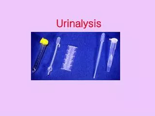
Performing the Urinalysis. Laboratory Procedures. Analyzing the Urine Sample. A complete urinalysis usually involves 3 steps. 1. Checking and recording the physical characteristics of the urine 2. Performing a chemical analysis using a multi-test dipstick

Performing the Urinalysis
E N D
Presentation Transcript
Performing the Urinalysis Laboratory Procedures
Analyzing the Urine Sample • A complete urinalysis usually involves 3 steps. • 1. Checking and recording the physical characteristics of the urine • 2. Performing a chemical analysis using a multi-test dipstick • 3. Centrifuging a small portion of the sample and examining the sediment under a microscope
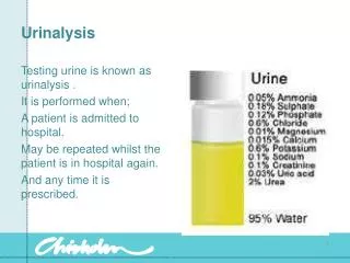
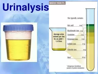
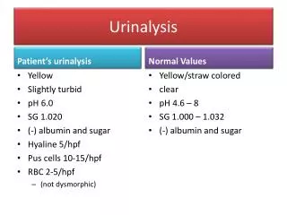
1. Checking and recording the physical properties of the urine • Physical properties include: • Color • Transparency • Odor • Specific gravity • Volume
2. Chemical Analysis • Many chemical tests can be performed on a small quantity of urine by using a dipstick • Each pad on dipstick is designed to test for a particular substance in the urine. • When the urine comes in contacts with the reagents, a chemical reaction will cause a color change based on the amount of the substance in the urine. • Color is compared to chart, and approximate amount of substance in urine can be determined
Chemical analysis • Certain drugs and medications may interfere with chemical tests. • Be sure to know any medications animal is currently receiving when performing a urinalysis.
Components on the Dipstick • Urine pH • Protein • Glucose • Ketones • Bilirubin • Urobilinogen • Blood • Nitrites
Urine pH • Number of how acidic or alkaline the urine is • pH is affected by diet. • Plant diets can cause alkaline urine • High protein diets can cause acidic urine • Small animal pH is usually acidic • In general dog and cat urine pH is 6-7 • Large animal urine is usually alkaline • Other factors may affect urine pH • Fever • Starvation • Certain drugs
Protein • Healthy animals will usually not have any protein in their urine although in some cases trace amounts can be found in concentrated dog and cat urine. • The urine protein level must be interpreted along with the Urine specific gravity. • Small amounts of protein are more significant in dilute or unconcentrated urine. • Protein may be lost in the urine due to glomerulopathies, inflammation, or hemorrhage.
Glucose • If the sugar in the blood is significantly higher than normal, some excess may be found in the urine. • Normal dog and cat urine should be negative for glucose. • In some cases if the urine is not run immediately, false glucose readings may occur.
Ketones • Ketones are the substances formed in the body during the breakdown of lipids. • Normal pet urine should be negative for ketones • When excess amounts of ketones are formed, their levels rise in the blood and then are released in the urine. • Can cause CNS depression and acidosis • May result in ketonuria caused by: • Starvation • Diabetes
Bilirubin • Pigment made by the liver from dead or dying red blood cells. • Small amounts may sometimes be found in healthy dogs. • Dogs can conjugate bilirubin in their kidneys, so small amount may be insignificant. • Bilirubin found in cat urine is a concern and can be a sign of liver disease, bile duct obstruction, or hemolysis.
Urobilinogen • Compound formed from bilirubin by intestinal tract. • Normal dogs and cats have small amounts of urobilinogen in their urine. • Results from dipstick are not considered very accurate and may be difficult to interpret. • Usually recorded as “normal” or “abnormal”.
Blood • Healthy pets may have a few red blood cells in their urine, but greater than ~5 cells per field may indicate a problem. • Hematuria can be due to a number of causes: • Trauma • Urinary Tract Infection (UTI) • Bladder Stones • Blood Clotting Problems
Nitrites • May be produced by the bacteria present in some infections • Test often shows a “false negative” and is considered inaccurate in pets. • However, if positive, should examine sediment closely for bacteria.
Examining Urine Sediment • After urine sediment is centrifuged (generally about 5 minutes), the top portion of the liquid is poured off and the sediment is resuspended and examined microsopically. • Indications for sediment exam include: • Provides additional information • A form of cytology • Must be interpreted with other clinical data, including physical and chemical composition of the urine.
Urine Sediment Exam Procedure • 1. Collect urine in a clean container • 2. Throughly mix specimen and transfer 3-5 ml volume to a centrifuge tube • 3. Centrifuge for 3-5 mins • 4. Pour off supernatant • 5. Leave approximately 0.5 ml of supernatant • 6. Resuspend urine sediment by tapping tube or flicking it w/ your finger. • 7. Examine a stained or unstained sediment. (Or both!)
Potential Sediment Elements • White blood cells • Red blood cells • Lipid droplets • Bacteria • Crystal • Casts
White blood cells • Larger than normal numbers of white blood cells may indicate inflammation from a bladder or kidney infection.
Lipid Droplets • Are common in the sediment of urine, especially in cats. • Number and size may vary • Should not be confused w/ air bubbles or RBC’s. They will be in focus when other structures are not.
Bacteria • Sediment is examined for presence of bacteria • Small amounts may be due to contamination, large amounts may indicate bladder infection (especially if sample is obtained via cystocentesis). • A urine culture and sensitivity may indicated with large amounts of bacteria.
Crystals • Made up of minerals and can sometimes be found in the urine. • Under certain conditions, crystals can clump together to form bladder stones (uroliths). • The pH of the urine may influence the type of crystal development • Some animals and species are more predisposed to crystal and stone formation.
Common Crystal Types • Struvite • Calcium Oxalate • Ammonium Urate
Casts (These will be covered in depth in Clin-Path!) • Urine Casts are small cylinder-shaped formations of cells and debris from inside the tubules of the kidneys • Presence and composition of casts can indicate kidney function • Types of casts: • Hyaline • Granular • Waxy • Fatty • Cellular • Mixed • Bile stained
Specific Gravity • Measures the concentrating ability of the kidney. • Done with a refractometer • There may be a spot on the dip-stick for SG, however in animals, these are not valid results.
Specific Gravity “normals” p. 158 Lab bk • “Normal” values in dogs is between 1.001-1.060 • “Normal” values in cats can range from 1.001 – 1.080 • There is no set “normal” value in animals, however the following guidelines should be used: • SG: below 1.008 is said to indicate dilution (hyposthenuric) • SG: 1.008-1.012 is said to be fixed or isosthenuric (same SG as plasma) • SG: 1.013 – 1.030 is considered normal if no dehydration suspected. • SG: above 1.025 implies renal tubule concentration ( in cats, this can indicate renal disease)