Download
1 / 45
450 likes | 521 Vues
Learn about the functions of the heart, lungs, and blood vessels in the circulatory system. Explore the mechanisms of blood flow, gas exchange, and pressure regulation in the body. Improve your knowledge of cardiovascular and respiratory physiology. |

E N D
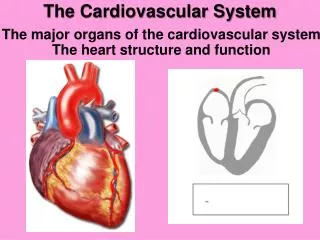
Figure 23.3B To lung To lung Left atrium Right atrium From lung From lung Semilunarvalve Semilunarvalve Atrioventricular(AV) valve Atrioventricular(AV) valve Right ventricle Left ventricle
Figure 23.4_s1 1 Diastole The semilunarvalves areclosed. The heartis relaxed. 0.4 sec The AV valves are open.
Figure 23.4_s2 2 1 Systole Diastole The semilunarvalves areclosed. The heartis relaxed. The atriacontract. 0.1 sec 0.4 sec The AV valves are open.
Figure 23.4_s3 2 3 1 Systole Diastole The semilunarvalves areclosed. The heartis relaxed. The atriacontract. 0.1 sec The ventriclescontract. 0.3 sec 0.4 sec The AV valves are open. The semilunarvalves are open. The AV valvesare closed.
Figure 23.9_s1 2 1 Typical bloodpressure:120 systolic70 diastolic Pressure in the cuffabove 120 Rubber cuffinflated with air 120 Artery Arteryclosed
Figure 23.9_s2 2 1 3 Typical bloodpressure:120 systolic70 diastolic Pressure in the cuffat 120 Pressure in the cuffabove 120 Rubber cuffinflated with air 120 120 Soundsaudiblein thestethoscope Artery Arteryclosed
Figure 23.9_s3 1 2 3 4 Typical bloodpressure:120 systolic70 diastolic Pressure in the cuffat 120 Pressure in the cuffabove 120 Pressure in the cuffat 70 Rubber cuffinflated with air 120 120 70 Soundsaudiblein thestethoscope Soundsstop Artery Arteryclosed
Figure 23.5A 3 1 2 4 Signals aredelayed at theAV node. Specialized musclefibers pass signalsto the heart apex. Signals spreadthroughout theventricles. Signals from theSA node spreadthrough the atria. SA node(pacemaker) Specializedmuscle fibers AV node Rightatrium Apex ECG
Figure 23.14A_s3 3 2 1 A platelet plugforms. A fibrin clotforms. Platelets adhere. Epithelium Connectivetissue Plateletplug Platelet Fibrin clot
Figure 23.7B Capillary Diffusion ofmolecules Interstitialfluid Tissuecell
Figure 23.11A Interstitial fluid Capillary wall Capillary lumen Nucleus ofepithelial cell Clefts betweenthe cells Muscle cell
Figure 23.11B Tissue cells Bloodpressure Osmoticpressure Venousend Arterialend Net fluid movementout of the capillary Interstitialfluid Fluid enters a lymph vessel
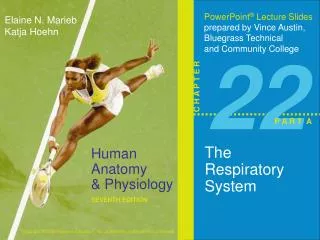
Figure 22.1 O2 Breathing 1 CO2 Lung Heart CirculatorySystem Bloodvessels Transportof gases bythe circulatorysystem 2 Capillary Mitochondria O2 Exchange ofgases withbody cells 3 CO2 Cell
Figure 22.11 Iron atom O2 loadedin lungs O2 O2 unloadedin tissues O2 Heme group Polypeptide chain
Figure 22.9_s3 Brain Cerebrospinalfluid 2 Breathing controlcenter respondsto the pH of bloodand cerebrospinal fluid. Nerve signalstrigger contractionof the rib musclesand diaphragm. 1 Medulla 3 Nerve signalsindicate CO2and O2 levels. CO2 and O2sensors in the aorta Heart Diaphragm Rib muscles