Download
1 / 7
80 likes | 340 Vues
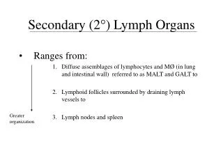
Secondary (2 °) Lymph Organs. Ranges from: Diffuse assemblages of lymphocytes and M Ø (in lung and intestinal wall) referred to as MALT and GALT to 2. Lymphoid follicles surrounded by draining lymph vessels to 3. Lymph nodes and spleen. Greater organization. Lymph nodes :.

E N D
Secondary (2°) Lymph Organs • Ranges from: • Diffuse assemblages of lymphocytes and MØ (in lung and intestinal wall) referred to as MALT and GALT to 2. Lymphoid follicles surrounded by draining lymph vessels to 3. Lymph nodes and spleen Greater organization
Lymph nodes: Afferent vessels Divided into 3 distinct regions: Cortex – w/ 1° follicles composed of APC’s Paracortex – w/T cells and DC’s w/ hi levels of MHC II Medulla – w/ plasma cells producing Ab’s Efferent vessel
Lymph nodes • Initial activation of B cells is thought to occur in paracortex • w/i 4-7 days, a few B cells and TH cells migrate to 1° follicles of cortex form 2° follicles
Spleen • Major role in IR’s to Ag’s in blood • Response center for systemic infections • Blood enters via splenic artery • Organ separated into regions of red pulp and white pulp
Spleen • Red pulp consists of sinuses w/ MØ’s, RBC’s site of RBC destruction/ removal • White pulp contains PALS w/ 1 follicles surrounded by lympho-cytes, MØ, and DC’s
Mucosal-associated lymphoid tissue (MALT) • Combined surface area of mucus membrane = 400 m2 (size of basketball court) • Major sites of entry • Protected by loose assemblage of organized lymphoid tissues (MALT) • Tonsils • Peyer’s patches
MALT (cont’d) • Each with groups of 30-40 lymphoid follicles