Download
1 / 69
690 likes | 1.12k Vues
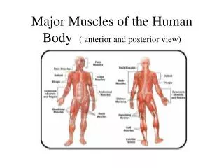
Memmler’s The Human Body in Health and Disease 11 th edition. Chapter 19 The Digestive System. Function and Design of the Digestive System. Chief functions…. we need NUTRIENTS ! Digestion Absorption Elimination Organs Digestive tract Accessory organs. Mucous membrane (mucosa)

E N D
Memmler’s The Human Body in Health and Disease11th edition Chapter 19 The Digestive System
Function and Design of the Digestive System • Chief functions….we need NUTRIENTS! • Digestion • Absorption • Elimination • Organs • Digestive tract • Accessory organs
Mucous membrane (mucosa) Digestive juice-secreting cells Mucus-secreting cells (goblet cells) Submucosa Connective tissue Contains blood vessels and nerves that help regulate digestion Smooth muscle Inner layer Outer layer Peristalsis Serous membrane Epithelium Loose connective tissue * Pic on pg. 410 – Figure 19.1 (same as next slide) The Wall of the Digestive Tract
Wall of the digestive tract. The mucous membrane of the small intestine shown here has numerous projections called villi. Zooming In: What type of tissue is between the submucosa and the serous membrane in the digestive tract wall?
The Peritoneum Membrane that lines the abdominopelvic cavity (shiny, thin that folds back to cover most organs) • Parietal peritoneum –outer portion of membrane • Visceral peritoneum – covers organs • Mesentery – double layered attached to posterior wall and small intestine • Mesocolon – from colon to posterior wall • Greater omentum – fatty layer from lower border of stomach to transverse colon • Lesser omentum – extends between stomach and liver
The Peritoneum (cont’d) Peritoneal cavity: space between two layers of the membrane • Greater peritoneal cavity: main portion, located in abdominal cavity, extends into pelvic cavity • Lesser peritoneal cavity: extends to the liver and the back attachment of the diaphragm
The abdominopelvic cavity. Subdivisions of the peritoneum fold over, supporting and separating individual organs. Zooming In: What part of the peritoneum is around the small intestine?
Organs of the Digestive Tract(muscular tube extending through the body) • Alimentary tract or gastrointestinal (GI) tract • Mouth • Pharynx • Esophagus • Stomach • Small intestine • Large intestine
The digestive system. Zooming In: What accessory organs of digestion secrete into the mouth?
The Mouth Also called oral cavity, processes food by • Ingestion • Mastication • Mixing with saliva • Deglutition (the tongue aids in swallowing and chewing and also aids in speech. has special sense receptors)
Deciduous teeth (baby teeth) Adult permanent teeth 32 teeth Incisors Cuspids (canines or eyeteeth) Molars Structure Dentin Blood vessels Nerves Gingiva Crown Enamel Roots Cementum The Teeth
The Pharynx Also called the throat • Oropharynx • Palatine tonsils • Nasopharynx • Laryngeal pharynx • Soft palate • Uvula • Epiglottis -in swallowing, the tongue pushes a bolus of food (chewed food) into the pharynx and the epiglottis closes over trachea
The Esophagus • Muscular tube (lubricated with mucous) • No digestion occurs here • Joins with stomach • Moves food by peristalsis • Esophageal hiatus – weakness in diaphragm causes hiatal hernia (pushes a portion through the space)
Question:What structure guards the entrance to the trachea during swallowing?a. uvulab. epiglottisc. esophageal sphincter
The Stomach – Structure • Additional angled muscle layer • Greater and lesser curvature • Fundus • Lower esophageal sphincter (LES) (cardiac sphincter) • Pylorus • Pyloric sphincter • Rugae
The Stomach – Functions • Store food and liquid • Secrete gastric juice (hydrochloric acid and pepsin) • Secrete mucus • Chyme: highly acidic mixture of gastric juice and food that leaves the stomach for the small intestine
Longitudinal section of the stomach. The stomach’s interior is visible, along with a portion of the esophagus and the duodenum. Zooming In: What additional muscle layer is in the wall of the stomach that is not found in the rest of the digestive tract?
Checkpoint 19-5:What type of food is digested in the stomach?
The Small Intestine – Structure • Duodenum • Jejunum • Ileum
The Small Intestine – Function • Secrete mucus • Secrete enzymes • Absorb digested food • Villi • Microvilli • Blood vessels • Specialized lymphatic capillaries (lacteals)
The small and large intestines. Zooming In: What part of the small intestine joins the cecum?
Checkpoint 19-6:What are the three divisions of the small intestine?Checkpoint 19-7:How does the small intestine function in the digestive process?
The Large Intestine — Structure • Cecum • Ileocecal valve • Vermiform appendix • Colon • Ascending • Transverse • Descending • Sigmoid • Rectum • Anal canal • Anus
The Large Intestine – Function • Secrete mucus • Reabsorb some water • Form feces (stool) • Defecation
Checkpoint 19-8:What are the divisions of the large intestine?Checkpoint 19-9:What are the functions of the large intestine?
Question:True or False?: The descending colon is a part of the small intestine.
Answer:False: The descending colon is part of the large intestine.
The Accessory Organs Release secretions through ducts into digestive tract • Salivary glands to mouth • All other organs to duodenum
Accessory organs of digestion. Zooming In:Into what part of the intestine do these accessory organs secrete?
The Salivary Glands • Functions of saliva • Moistens food • Facilitates mastication and deglutition • Helps keep teeth and mouth clean • Production of saliva • Parotid glands • Submandibular (submaxillary) glands • Sublingual glands
The Liver – Structure • Right, left lobes • Portal vein • Hepatic artery
The Liver – Function • Manufacture bile • Store glycogen, convert to glucose • Modify fats • Store vitamins, iron • Form blood plasma proteins • Destroy old red blood cells • Synthesize urea • Detoxify harmful substances
The Gallbladder Bile • Flows from liver through cystic duct • Is stored in gallbladder • Flows through cystic duct and common bile duct to duodenum when needed
The Pancreas • Releases enzymes that digest fats, proteins, carbohydrates, and nucleic acids • Produces alkaline (basic) fluid to neutralize acidic chyme in small intestine • Produces insulin and glucagon to regulate sugar metabolism
Checkpoint 19-11:What is the role of the gallbladder?Checkpoint 19-12:What is the role of the bile in digestion?
Question: What two organs are involved in the production of bile?a. the gallbladder and the stomachb. the liver and the gallbladderc. the liver and the spleen
Enzymes and the Digestive Process Enzymes • Speed up rate of chemical reactions • Are not changed or used up in reactions • Are proteins • Are highly specific in their actions
The Role of Water Process of digestion technically hydrolysis (split by water) Water is used to • Produce digestive juices • Dilute food • Aid chemical process of digestion • Added to nutrient molecules as they are split by enzymes
Digestion, Step-by-Step • Mouth • Chews food, mixes with saliva • Some starches changed to sugars • Stomach • Secretes hydrochloric acid, enzymes • Secretes mucus • Forms chyme • Begins digestion of proteins (with pepsin) • Small intestine • Mixes chyme with bile • Receives pancreatic juice enzymes • Produces enzymes • Emulsifies fats
Checkpoint 19-13:What organ produces the most complete digestive secretions?
Absorption • Villi in mucosa of small intestine • Arteriole and venule bridged with capillaries • Capillaries absorb • Simple sugars • Small proteins • Amino acids • Simple fatty acids • Water • Portal system transports nutrients to liver
Absorption of Fats • Lacteals absorb fat • Fat/lymph mixture (chyle) drains from small intestine • Chyle merges with lymphatic circulation, enters blood in veins near heart • Liver further processes absorbed fats
Absorption of Vitamins and Minerals When vitamins and minerals are • Dissolved in water • Absorbed directly into blood • Incorporated in fats • Absorbed with fats • Produced by bacterial action • Absorbed in large intestine
Control of Digestion • Nervous • Parasympathetic stimulation increases activity • Sympathetic stimulation decreases activity • Hormonal • Digestive organs produce hormones • Gastrin • Gastric-inhibitory peptide (GIP) • Secretin • Cholecystokinin
Checkpoint 19-15:What are the two types of control over the digestive process?
Hunger and Appetite • Hunger • Hypothalamic centers regulate • Blood nutrient levels stimulate • Satisfied by adequate meal • Appetite • No relationship to need for food • May not be satisfied by adequate meal • Leptin; hormone produced in adipose tissue; involved in weight regulation