Lupus in Pregnancy
Lupus in Pregnancy. Darren Farley, MD Clinical Assistant Professor Division of Maternal-Fetal Medicine Dept. of Obstetrics and Gynecology University of Kansas School of Medicine – Wichita. No financial interests to disclose. Objectives. Physiologic changes Lupus overview

Lupus in Pregnancy
E N D
Presentation Transcript
Lupus in Pregnancy Darren Farley, MD Clinical Assistant Professor Division of Maternal-Fetal Medicine Dept. of Obstetrics and Gynecology University of Kansas School of Medicine – Wichita
Objectives • Physiologic changes • Lupus overview • Care in pregnancy • Congenital heart block • Hydroxychloroquine • Lupus flares
Maternal Physiology Robinson, 2012
Maternal Physiology • Estrogens upregulate T-cell responses, immunoglobulin synthesis, and leukocyte production of IL 1, 2, 6, TNFα • Cell-mediated immunity is depressed • Decreased ratio of T cells to B cells • Increased ratio of suppressor T cells to helper T cells • Decreased ratio of lymphocytes to monocytes • Inhibition of complement activation in the placenta may be essential for fetal survival • Trophoblast may be a target of autoimmunity. Creasy
Maternal Physiology • AFP suppresses lymphocyte function • IL-1, IL-3, TNF-α, IFN γ, Granulocyte-macrophage colony-stimulating factor are critical in sustaining pregnancy. • IL-3 levels - low in women with RPL • Pregnancy - Total C3, C4, and hemolytic (CH50) complement levels – unchanged / increased • Increase in classic pathway complement activation • Complement activation can result in excess soluble vascular endothelial growth factor receptor type 1 (sFlt-1), which has implications for placental development and the risk for preeclampsia. Creasy
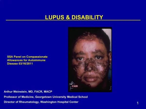
SLE • Chronic autoimmune d/o with disease flares and remissions • Can affect all organs • Mild cases – skin, musculoskeletal system • More severe – kidney, brain • Possible manifestations are arthralgias, rashes, renal abnormalities, neurologic complications, thromboemboli, myocarditis, serositis
Epidemiology • Prevalence of lupus varies with population • 5-125/100,000 people • Affects 1% of pregnancies • Lifetime risk of developing lupus is 1/700 • peaks at 30 y/o • Women : Men – 9:1 • Ethnic groups • African Americans • Hispanics
Etiology • Unknown • Genetic linkage • 5-12% of affected individuals have an affected relative • 25-50% of monozygotic twins are concordant for the disease • Alterations in HLA system • HLA-B8, HLA-DR3, HLA-DR2 • Abnormal B and T cell biology and immune clearance mechanisms
Pathophysiology • Damage due to immune complex deposition, complement activation, inflammation, fibrosis • Renal, MSK, hepatic, platelets • Autoantibodies • Antinuclear antibody – most common, ‘lab is open’ • Increased in pregnancy – 10% of asymptomatic pregnant women without autoimmune disease have ANA ab compared to 2% of nonpregnant controls • Screening for lupus b/c of high prevalence in gen pop • Anti - dsDNA antibody and anti-Smith Ab - more specific for lupus; dsdna ab correlates with disease activity • Antiphospholipid antibodies • Anti – Ro/SSA, anti- La/SSB more often associated with sjogrens sd, but seen in 20-40% of females with lupus • Associated with neonatal lupus syndrome
Symptom frequency • Fatigue – 80-100% • Fever – 80-100% • Arthritis – 95% • Myalgia – 70% • Weight loss – 60% • Photosensitivity – 60% • Malar rash - 50% • Nephritis – 50% • Pleurisy – 50% • Lymphadenopathy – 50% • Pericarditis – 30% • Neuropsychiatric – 20%
Criteria for Diagnosis • Per American College of Rheumatology • Need 4 of 11 (serially or at one time) • Malar rash (erythema over malar eminences) • Discoid rash (erythematous raised patches) • Photosensitivity (unusual rxn to sunlight) • Oral ulcers (oral, nasopharyngeal) • Arthritis (nonerosive, 2+ peripheral joints) • Serositis (pleuritis, pericarditis) • Nephritis (>500mg/d proteinuria or cellular casts) • Neurologic disorder (seizures, psychosis, stroke with other causes r/o) • Hematologic disorder (hemolytic anemia with reticulocytosis, thrombocytopenia <100k, leukopenia <4000 2 occasions, lymphopenia <1500 2 occasions) • Immunologic disorder (anti-dsDNA, anti-Sm, positive LAC ACA, false pos RPR or other serologic test for syphilis for 6 months confirmed by treponema pallidum immobilization or fluorescent treponemal ab absorption test) • Antinuclear antibodies (without being on drugs associated with drug induced lupus syndrome • <4 of 11 = lupus-like syndrome
General Morbidity/Mortality • Renal and cardiovascular disease • Thrombosis • Infection • Survival rates • 5y – 93% • 10y – 85% • 15y – 79% • 20 y – 68% • Risk factors for death from lupus • Lupus nephritis • Thrombocytopenia • Lung involvement • High disease activity at time of diagnosis
Pregnancy Outcomes • Effect of SLE on pregnancy • Increased stillbirth rate 25x (150/1000) • Esp with antiphospholipid antibodies • Increased preeclampsia rate to 20-30% (7-10%) • Increased IUGR rate to 12-32% • Increased preterm delivery rate to 50-60% (12-15%) • Increased PPROM rate • Neonatal lupus only in Ro/La antibody positive patients • Effect of pregnancy on SLE • Worsening renal status if nephropathy (Cr 1.5) present • Increased flares if active disease at start of pregnancy
Chronic Renal Insufficiency and Pregnancy Outcomes (vs serum creatinine mg/dL) Queenan 2007
Renal Biopsies in Pregnancy • Complications - • Hematoma formation • Bleeding • Death • Usually do not help change management • If steroids are the change in management, then steroid administration is of lower risk than a renal biopsy
Goals of Management • Disease control/remission before pregnancy • Avoid drugs that harm the fetus • Prompt detection of preeclampsia and placental insufficiency • Discern between lupus exacerbations and preeclampsia • Appropriate detection and treatment of lupus flares
Preconception counseling • Potential complications – preeclampsia, preterm labor, miscarriage, fetal death, fetal growth restriction, and neonatal lupus • Evaluate lupus activity – delay pregnancy until remission • Evaluate for nephritis (24 hr urine), hematologic abnormalities (CBC), antiphospholipid abnormalities • Discontinue NSAIDS and cytotoxic agents
NSAIDS • NSAIDS – inhibits cyclooxygenase, lipoxygenase, reduces prostaglandin synthesis • Class D • Avoid especially in 3rd trimester • Cross placenta, blocks prostaglandin synthesis in fetal tissue • Premature closure of ductus arteriosis, fetal pulmonary hypertension, NEC, fetal renal insufficiency • Occurs with selective COX-II inhibitors • ASA crosses placenta and can affect fetal platelet function and is associated with intracranial fetal hemorrhage in 3rd trimester; avoid in pregnancy • Used outside of pregnancy – most common anti-inflammatory agent
Hydroxychloroquine • Hydroxychloroquine (antimalarial/antirheumatic; binds DNA, interferes with vesicle functions, inhibits phospholipid metabolism; immunosuppressive by inhibiting rheumatoid factor, acute phase reactants, enzymes) • Stopping this in pregnancy is associated with increased risk of lupus flares, continuing this drug is recommended if needed to control lupus (prospective study by cortes-hernandez showed the increased risk) • Large series show no increased risk of anomalies • Used in prevention of malaria with increase of fetal anomalies • Not associated with increased r/o fetal malformations • Class C • Chloroquine possible teratogenic in initial studies • Ototoxicity, eye development Buchanan, 1996; Khamashta 1996 Klinger 2001; Motta 2002
Glucocorticoids • Glucocorticoids (antiinflammatory, glucocorticoid, mineralocorticoid) • Preg class C • Avoid fluorinated glucocorticoids b/c they cross the placenta • Hydrocortisone, prednisone, prednisolone inactivated by 11-beta hydroxysteroid dehydrogenase in the placenta allowing <10% of active drug to reach fetus • High dose associated with maternal/fetal A/E • Osteoporosis (tx with vit D, ca2+); glucose intolerance, sodium, h2o retention; hypertension, infection; avascular necrosis • Preg complications – GDM, preeclampsia, PPROM, IUGR • Incidence of fetal adrenal suppression with maternal tx is low • Avoid empiric treatment, use at lowest possible dose • Stress dose steroids (hydrocortisone 100mg IV q8hr in labor and for 24 hr PP) • Use if chronic steroids (>5mg/day for >2-4 weeks prior to delivery)
Azathioprine • Azathioprine (inhibits T lymphocytes) • Class D • Teratogenic in animals, appears safe in humans • Associated with IUGR • Neonatal immunosuppression • Indicated in pregnancy if chronic high doses of steroids is not controlling symptoms or to lower steroid dose
Cyclophosphamide • Cyclophosphamide (alkylates and cross links DNA) • Preg class D • Cleft palate, skeletal abnormalities, abnormal renal function • Avoid, esp in first trimester • May be needed in cases of severe proliferative nephritis (drug of choice in nonpregnant patients with proliferative lupus nephritis) • Crosses placenta
Methotrexate • Methotrexate (inhibits dihydrofolate reductase; inhibits lymphocyte proliferation) (folate antagonist) • Preg class X • Avoid • Embryolethal, IUFD • Congenital anomalies
Cyclosporine • Cyclosporine A (inhibits T lymphocytes) • Preg class C • Data comes from use in renal transplant patients, not an animal teratogen, appears safe in humans, long term follow up studies are limited
Tacrolimus • Tacrolimus (inhibits T lymphocyte activation, immunosuppressant) • Dose in liver transplant • 0.1-0.15mg/kg/d po divide q12 hr • Preg class C • Therapeutic drug levels 5-20 ng/ml just before next dose; time to steady state 3 days • Monitor creatinine, K, fasting blood glucose, serum drug levels
Pregnancy - FDA classes • A – controlled studies show no fetal risk in any trimester, probability of fetal harm is remote • B – animal studies, no risk; if risk in animal studies, controlled human studies do not confirm harm • C – harm in animal studies with no controlled human studies; no available human or animal studies • D – human studies show fetal risk but r/b relative to medical state of mother may support use • X – animal/human studies show fetal risk or abnormalities, use is contraindicated during pregnancy or in women who may become pregnant
Pregnancy • Labs/Evaluation • *CBC, CMP, 24hr UA for TP/CC • *Antiphospholipid antibodies • *Anti-Ro and anti-La antibodies • *Anti-dsDNA antibody • Complement (C3 and C4 or CH50) • Monthly CBC, Platelet count, Complement and anti-dsDNA antibody • Maternal echocardiogram if disease present >3-5 yrs, cardiac complications, associated CHTN, lupus nephritis
Lupus and Presence of Antiphospholipid Antibodies • 1/3 of lupus patients • Risks – thrombosis, fetal loss • + APA and history of fetal loss = APLS • Heparin/lovenox is recommended • Thromboprophylaxis • Due to increased thrombosis risk • Data is lacking that reveals improved outcomes (less SABs, IUFDs, etc) unless APLS is diagnosed
Antenatal care • Frequent visits to assess lupus status, screen for hypertension • Continue hydroxychloroquine • Depends on control as to whether to initiate it if patient is not medicated • Monitor for exacerbations/flares • If chronic hypertension – monitor as such • APLS – see above
Antenatal care • Between 18 and 25 weeks (mothers with anti-ro/la antibodies) • *Screening fetal echocardiogram • Fetal electrocardiogram through echocardiography • Vs. *weekly FHR checks • +/- Dexamethasone • Serial ultrasounds to evaluate fetal growth • Antenatal surveillance at 32 weeks or earlier if indicated
Neonatal Lupus • Rash, thrombocytopenia, hepatitis, hemolytic anemia • Transient • Complete heart block – Permanent • Only if +SSA/B antibodies • 25% risk of rash (recurrence risk 25%) • <3% risk of heart block (RR 18%)
Congenital Heart Block • SSA/B + increases risk • Prior history of child with heart block increases risk • Dexamethasone – limited data that shows clear benefit • Hydroxychloroquine Izmirly, 2010
Fetal PR Interval Wojakowski 2009
PRIDE Study • 127 women evaluated, 95 completed course – all had Ro or La Ab • Fetal echo, weekly 16-26 weeks • PR >150msec – 1st degree • 92 - Normal PR intervals • 3 with complete heart block, without prolonged PR interval preceding it • Tricuspid regurgitation, atrial echodensity • 2 had PR intervals >150msec, 22 weeks • Dexamethasone initiated, reported to prevent progression and resolved the 1st degree • Recurrence – 19% with previous heart block • 3% without previous heart block
Evaluation Of The Risk Of Anti-ssa/Ro-ssb/La Antibody-associated Cardiac Manifestations Of Neonatal Lupus In Fetuses Of Mothers With Systemic Lupus Erythematosus Exposed To Hydroxychloroquine • TLR signaling in pathogenesis of neonatal heart block • Hydroxychloroquine is a TLR inhibitor • A TLR inhibitor, might reduce the risk of anti-SSA/Ro/SSB/La antibody associated cardiac manifestations of NL • Cardiac-NL children (N=50) and controls (N=151) were drawn from the following overlapping pregnancy studies: Research Registry for NL; PR Interval and Dexamethasone Evaluation in Cardiac-NL; and Predictors of Pregnancy Outcomes: Biomarkers in Antiphospholipid Syndrome and SLE • Ro/La +; SLE dx • Results Seven (14%) of the cardiac-NL children were exposed to HCQ compared with 56 (37%) of the controls (p=0.002; OR 0.28; 95% CI 0.12 to 0.63). • Concluded – in mothers with SLE with anti-SSA/Ro/SSB/La antibodies, exposure to HCQ during pregnancy may decrease the risk of fetal development of cardiac-NL Izmirly, 2010
Maternal Autoantibody Levels In Congenital Heart Block And Potential Prophylaxis With Antiinflammatory Agents • Retrospective, 2007-2011, Ro/La + • N =33 • Higher Anti La titers in pregnancies c/b heart block; no difference if Ro Ab • Did not have to have SLE, etc • 94% of fetuses maintained normal conduction when the mother was treated with hydroxychloroquine or daily prednisone therapy throughout pregnancy, compared to 59% in the untreated group (odds ratio, 0.1; P = .04). • Maternal treatment with either hydroxychloroquine or daily low-dose prednisone throughout pregnancy may provide a protective effect. Tunks 2013
Lupus Flares • Incidence in pregnancy 15-63% • Studies support and refute that pregnancy increases the incidence of flares • Risk factors • Active disease at conception (50% vs 20%) • Active nephritis • Abrupt discontinuation of hydrochloroquine
Symptoms Ratigue, fever, arthralgias/myalgias, weight loss, rash, renal deterioation, serositis, LAD, CNS symptoms Titers of antibodies Rising titers of dsDNA Ab with falling complement levels suggest impending flare Diagnosis Of Flare
Lupus Vs Preeclampsia • Lupus flare • Arthritis, leukopenia, thrombocytopenia, rashes, pleuritis, fevers • Htn, proteinuria, coagulopathy possible • Rising antidsdna titer, active urinary sediment, low complement levels suggest lupus flare • Complement levels (C3, C4, CH50) generally rise in pregnancy and are unaffected by uncomplicated preeclampsia • Normal uric acid • Differentiation near term likely not worthwhile, deliver for suspected preeclampsia and initiate tx for lupus flare if patient does not get better
Lupus Flare vs. Preeclampsia Foley OB ICU care manual
Renal Biopsies in Pregnancy • Complications - • Hematoma formation • Bleeding • Death • Usually do not help change management • If steroids are the change in management, then steroid administration is of lower risk than a renal biopsy
Renal Biopsy • Risks in pregnancy • Hematoma formation • Renal dysfunction • Lupus Nephritis • Increased mesangial matrix and mesangial hypercellularity (increased leukocytes) • Preeclampsia - Endotheliosis