Download
1 / 42
430 likes | 884 Vues
Motor Functions of the Spinal Cord and the Cord Reflexes. Prof. Dr. Bayram Yılmaz Yeditepe University Faculty of Medicine Department of Physiology. GENERAL FEATURES OF THE SPINAL CORD. The spinal cord:. is continuous with the brain mediates spinal reflexes

E N D
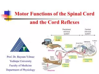
Motor Functions of the Spinal Cord and the Cord Reflexes Prof. Dr. Bayram Yılmaz Yeditepe University Faculty of Medicine Department of Physiology
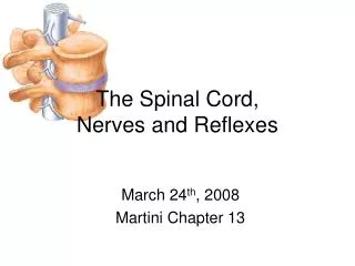
GENERAL FEATURES OF THE SPINAL CORD The spinal cord: is continuous with the brain mediates spinal reflexes is a site for nervous integration (contains synapses) provides pathways to and from the brain
PROTECTION AND COVERINGS Vertebral canal Meninges Cerebrospinal fluid
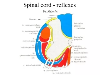
INTERNAL ANATOMY OF THE SPINAL CORD Gray matter gray commissure dorsal horn white matter nuclei horns gray matter lateral horn dorsal – sensory ventral – motor lateral -- autonomic ventral horn White matter Gray commissure Central canal central canal
SPINAL NERVE ROOTS Dorsal root ganglion – cell bodies of sensory neurons Dorsal root – axons of sensory neurons Ventral root – axons of motor neurons dorsal root ganglion dorsal roots dorsal root ganglion ventral roots
Spinal Animal and Decerebrate Animal • Spinal animal: the spinal cord is transected in the neck, most of the cord still remains functional • Most spinal functions are severely depressed below the level of transection • Decerebrate animal: the brain stem is transected in the middle to lower part of the mesencephalon
Decerebrate Rigidity • Decerebrate Rigidity: transection of the brainstem at midbrain level (above vestibular nuclei and below red nucleus) • Symptoms include: • extensor rigidity or posturing in both upper and lower limbs
Decerebrate Rigidity Results from: • loss of input from inhibitory medullary reticular formation (activity of this center is dependent on input from higher centers). • active facilitation from reticular formation (intrinsically active, and receives afferent input from spinal cord).
Organisation of the Spinal Cord for Motor Functions • The cord gray matter is the integrative area for the cord reflexes • Sensory transmission to • Gray matter of the cord • Higher centers of the nervous system http://msjensen.cehd.umn.edu/1135/Links/Animations/Flash/0016-swf_reflex_arc.swf
Anterior Motor Neurons • Alpha motor neurons: (Aa) • Stimulation of a single alpha motor fiber excites anywhere from three to several hundred skeletal muscle fibers • Gamma motor neurons: (Ag) • Stimulate small, special skeletal muscle fibers called intrafusal fibers • These fibers constitute the middle of the muscle spindle
The extrafusal muscle fibers are innervated by Alpha motor neuron The intrafusal muscle fibers are innervated by Gamma motor neurons
A motor unit is a single motor neuron (a motor) and all (extrafusal) muscle fibers it innervates Motor units are the physiological functional unit in muscle (not the cell) All cells in motor unit contract synchronously Motor units
Motor units and innervation ratio Innervation ratio Fibers per motor neuron Extraocular muscle 3:1 Gastrocnemius 2000:1
Interneurons • They are about 30 times more than the motor neurons • These cells are highly excitable (capable of firing rate at 1500 times per second) • Divergence, convergence and repetetive discharge • Only a few incoming sensory or brain signals terminate directly on the anterior motor neurons • Interconnections
Renshaw Cell Inhibitory System • There are a large number of neurons called Renshaw cells • They send inhibitory signals to surrounding motor neurons • Function in lateral inhibition • Propriospinal fibers: • Multisegmental connections from one spinal cord level to other levels • These ascending and descending propriospinal fibers provide pathways for multisegmental reflexes
Muscle Sensory Receptors • Muscle spindles • Golgi tendon organs
Muscle Spindles • Intrafusal fibers are very small skeletal muscle fibers • Central region of these fibers do not contain actin or myosin filaments (this portion does not contract) • The end portions that contract are excited by gamma motor (efferent) fibers • Sensory information from muscle spindle: • Lenghthening the whole muscle stretches the midportion of the receptor • Contraction of the end portions of intrafusal fibers stretches the midportion of the spindle
Primary and Secondary Endings • Primary ending (or annulospiral ending): In the center of the receptor area, a large sensory nerve fiber encircles the central portion of each intrafusal fiber • Secondary ending: innervates the receptor region on one or both sides of the primary ending
Static and Dynamic Responses of the Receptor • Static response: When the muscle spindle is stretched slowly, impulses from both primary and secondary endings increase in proportion to stretching • Dynamic response: Response of the primary ending (but not the secondary ending) to rate of change of receptor length • Continuous discharge of the muscle spindle under normal conditions • Positive or negative signals
Muscle Stretch Reflex • The simplest manifestation of muscle spindle function is the muscle stretch reflex
Exciting a muscle spindle occurs in two ways Applying a force that lengthens the entire muscle Activating the motor neurons that stimulate the distal ends of the intrafusal fibers to contact, thus stretching the mid-portion of the spindle (internal stretch) The Stretch Reflex
The Stretch Reflex • Whatever the stimulus, when the spindles are activated • their associated sensory neurons transmit impulses at a higher frequency to the spinal cord http://sites.sinauer.com/neuroscience5e/animations16.01.html
Dynamic and Static Stretch Reflexes • The dynamic stretch reflex occurs within a fraction of a second after the muscle has been stretched • Then a weaker static reflex continues for a prolonged period thereafter • “Damping” function of the dynamic and static stretch reflexes • This is also called signal averaging function of the muscle spindle reflex which makes the contraction relatively smooth
Role of muscle spindle in voluntary motor activity • About 31% of all motor fibers to the muscle are small type gamma efferent fibers • Coactivation of alpha and gamma motor fibers, and excitation of extra- and intra-fusal muscle fibers • This coactivation keeps the muscle spindle reflex from opposing the muscle contraction • Also, it maintains the proper damping function of the muscle spindle, regardless of any change in muscle length
Brain Areas for control of gamma motor system • Gamma efferent system is excited specifically by signals from bulboreticular facilitatory region of the brain stem • Cerebellum, basal ganglia, the cerebral cortex • Muscle spindle system stabilizes body position during tense action
Clinical Applications of the Stretch Reflex • Knee jerk and other muscle jerks http://www.brainviews.com/abFiles/AniPatellar.htm
Golgi Tendon Reflex • Golgi tendon organ helps control muscle tension • The tendon organ has both dynamic and static responses
Transmission of Impulses from Golgi Tendon Organ to the CNS • Signals from tendon organs are transmitted by Ib fibers • Transmit signals to the local areas of the cord • After synapsing in the dorsal horn, spinocerebellar tracts project to cerebellum • Other tracts to cerebral cortex • The local cord signal excites a single inhibitory interneuron that inhibits the individual motor neuron • Inhibitory nature of the tendon reflex & its significance
Flexor Reflex and Withdrawal Reflex • Withdrawal of the extremity from the stimulating object is called flexor reflex • Flexor reflex and stimulation by nociceptors • The pathways for eliciting a flexor reflex don’t pass directly to the motor neurons, instead pass first into a neuronal pool • The shortest possible circuit is a three or four neuron pathway
Flexor Reflex and Withdrawal Reflex • 1) Diverging circuits to spread the reflex to the necessary muscles for withdrawal • 2) Circuits to inhibit the antagonist muscles (reciprocal inhibition circuits) • 3) Circuits to cause after-discharge lasting many fractions of a second after the stimulus is over
Flexor Reflex and Withdrawal Reflex http://www.sumanasinc.com/webcontent/animations/content/reflexarcs.html
Crossed Extensor Reflex • About 0.2 to 0.5 second after a stimulus elicits a flexor reflex in one limb, the opposite limb begins to extend
Reciprocal Inhibition and Reciprocal Innervation • Inhibition of the antagonistic muscles is called reciprocal inhibition • In essence, the stretch stimulus causes the antagonists to relax so that they cannot resist the shortening of the “stretched” muscle caused by the main reflex arc
Reflexes of Posture and Locomotion • Cord “righting” reflex of a spinal animal • Stepping and walking movements • Rhytmical stepping movements are frequently observed in the limbs of a spinal animal • This oscillation back and forth between flexor and extensor muscles can occur even after the sensory nerves have been cut • Diagonal stepping of all four limbs (Mark Time Reflex) http://bcs.whfreeman.com/thelifewire/content/chp46/46020.html
Scratch Reflex • Scratching reflex involves two functions: • A position sense that allows to find the exact point of irritation on the surface of the body • A to- and- fro scratching movement
Spinal Cord Reflexes that Cause Muscle Spasm • Muscle spasm resulting from a broken bone • Abdominal muscle spasm in peritonitis • Muscle cramps • Severe cold • Lack of blood supply • Over-exercise
Autonomic Reflexes in the Spinal Cord • Many types of segmental autonomic reflexes are integrated in the cord • Changes in vascular tone resulting from changes in local skin heat • Sweating • Intestino-intestinal reflexes and some motor functions of the gut • Peritoneo-intestinal reflexes that inhibit GI motility • Evacuation reflexes for emptying the full bladder
Spinal Cord Transection and Spinal Shock • When the spinal cord is suddenly transected in the upper neck, all cord functions including the cord reflexes become depressed (spinal shock) • After a few hours to a few weeks, the spinal neurons gradually regain their excitability • At the onset of the spinal shock, arterial pressure falls dramatically (as low as 40 mmHg) • Depressed skeletal muscle reflexes • Sacral reflexes for control of bladder and colon are suppressed.