Download
1 / 92
1.01k likes | 1.54k Vues
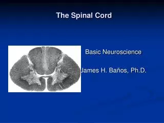
Motor Functions of the Spinal Cord. BIEN 500 Steven A. Jones. Spinal Cord Function. Conduct signals to the periphery of the body. Process these signals as necessary Reflex actions More complicated actions, such as walking. Spinal Cord Divisions. Anterior Motor Neurons

E N D
Motor Functions of the Spinal Cord BIEN 500 Steven A. Jones
Spinal Cord Function • Conduct signals to the periphery of the body. • Process these signals as necessary • Reflex actions • More complicated actions, such as walking
Spinal Cord Divisions • Anterior Motor Neurons • Alpha Motor Neurons (large skeletal muscles) • Type A nerve fibers • Enervate large skeletal muscles • Stimulate 3 to several hundred fibers each • Gamma Motor Neurons • Type A nerve fibers • Send signals to muscle spindles • Interneurons • Perform Integrative functions • Most signals in spine are processed through these • Renshaw Cell Inhibitory System • Suppresses spread of signals • Located in the ventral horn
Motor Spindles • Stimulated by stretching • Send signals as a result of: • Change in length of muscle. • Change in tension on the muslce. • Primary ending • Group Ia fiber • Important for sensing dynamic changes • Secondary ending • Group II fiber • With Primary ending, sends a static response
Muscle Stretch Reflex • Muscle stretch causes contraction • First dynamic (short term), then static (long term) • Shortening of muscle will cause inhibition Anterior Horn Motor Nerve Proprioceptor Nerve (Ia) Muscle Spindle Posterior Horn
Feedback Function of Spindle This effect is not surprising, is it? Force of Contraction With Spindle Without Spindle 3 0 Time (s) 31% of the motor nerve fibers are gamma efferent fibers
Gamma Motor System • Excited by bulboreticular facilitory region of the brain stem via: • Cerebellum • Basal ganglia (in the brain) • Caudate nucleus • Putamen • Globus pallidus • Substantia nigra subthalamic nucleus • Crebral cortex
Knee Jerk and Clonus Muscle Length Clonus (think time delay) E.g. fatigue when standing on tip toes. Knee Jerk 400 0 Time (ms)
Golgi Tendon Organ • Detects tension rather than length. • Found between tendon and muscle. • Signals transmitted by type Ib nerve fibers. • Negative feedback to prevent overstimulation. • May help even out muscle load on fibers. Tendon Golgi Tendon Organ Muscle
Flexor/Withdrawal Reflex • Also called nociceptive reflex • Pain initiates flexion, without passing through brain • Passes through interneurons • Get extension in opposite limb
Flexor Reflex Extensor Inhibition Extensor Stimulation Flexor Stimulation Flexor Inhibition Interneurons Pain Signal from Hand Reciprocal Inhibition: Stimulus exciting one muscle will inhibit the antagonist muscle.
Spinal Cord Reflexes • These responses occur in decerebrate animlas. • Walking • Galloping • Scratching
Muscle Spasm • Broken bone • Abdominal muscle spasm • Muscle cramps • Severe cold • Lack of blood flow • Overexercise • Contraction may stimulate more contraction – positive feedback.
Autonomic Spinal Cord Reflexes • Vascular tone (e.g. change in skin perfusion) • Sweating • Intestinal reflexes • Control of gut muscles • Inhibition of gastrointenstinal motility (e.g. irritation) • Bladder/Bowel control
Spinal Shock • Occurs when spinal cord is deprived of brain input. • Spine recovers after days. • Similar mechanism applies to most nerves • Recovery may be excessive • Affected functions: • Arterial blood pressure • Skeletal muscle reflexes are blocked • Bladder/bowel control are lost (but recover)
Cortical and Brain Stem Control of Motor Function BIEN 500 Steven A. Jones
The Cerebellum, the Basal Ganglia, and Overall Motor Control BIEN 500 Steven A. Jones
Organization of the Brain Thalamus Cerebellum Regions of the Basal Ganglia
Motor Functions of the Cerebellum • Running • Playing a musical instrument • Talking • Writing • Typing • I.e. Repeated, learned actions.
Mechanism of Action of Cerebellum • Sequences motor activities • Monitors and corrects motor activities to conform to signals from the motor cortex & other parts of the brain. • A negative feedback system.
Anatomy Anterior Lobe • Afferent – Incoming • From brain • From periphery • Efferent – Outgoing • From brain • From periphery Posterior Lobe Flocculonnodular Lobe Oldest, Vestibular (equilibrium) control
The Purkinje Cell Molecular Layer Billions of Parallel Nerve Fibers Purkinje (Inhibitory) Constantly firing Purkinje Layer Granule Layer Granule Cells Deep Nuclei Input from Inferior Olive (Climbing Fiber) strong stimuli Input from Elsewhere (Mossy Fiber) weak stimuli Deep Nuclear Cell Constantly firing Output Multiply by 30,000,000
Learning (Perkinje Cells) • Inferior olivary complex receives intent and execution information – sends error signal. • Climbing fiber rate – 1 per second • Mismatch between desired movement & actual movement increases/decreases rate. • Firing rate changes sensitivity of perkinje cells. • Error signal is then stopped.
Feedback Control of the Distal Limb Motor Cortex Thalamus Red Nucleus Feedback from Cerebellum Intermediate zone of cerebellum Intended Movements Actual Movements Muscles Generally important for movements that are too fast to be corrected on the fly.
Effects of Cerebellar Damage • Slow development of movements • Weak development of force • Overshoot of motion Thus, the cerebellum functions to plan, sequence and time complex motions.
Non-Motor Functions of the Cerebellum • Estimation of speed. • Prediction of timing. • Interpreting spatiotemporal relations.
Abnormalities of the Cerebellum • Dysmetria and Ataxia/Past Pointing • Discoordinated movements/Overshoot • Dysdiadochokinesia – don’t know where your body parts are in fast motion. • Dysarthria – cannot coordinate speech • Intention tremor – movement overshoot • Cerebellar nystagmus – tremor of the eyeballs • Hypotonia – loss of muscle tone.
Basal Ganglia • Include the following components of the brain: • Caudate nucleus • Putamen • Globus pallidus • Substantia nigra • Subthalamic nucleus • Located lateral to the thalamus • Nearly all motor & sensory nerves from the crerebral cortex pass between the caudate nucleus and putamen.
Basal Ganglia Thalamus Amygdala Putamen and Globus Palidus Cerebellum Caudate Nucleus
Basal Ganglia • Control complex motor activity • E.g. writing letters of the alphabet • Cutting paper with scissors • Shooting a basketball • Hammering nails • Throwing a football/baseball
Parkinson’s Disease • Destruction of the pars compacta in the substantia nigra • Prevents activity of dopamine-secreting nerve fibers to the caudate nucleus. • Dopamine = Inhibitor • Causes • Rigidity of musculature • Involuntary tremor (3-6 Hz) • Difficulty initiating movements
Treatment for Parkinson’s • L-Dopa • Gets converted to dopamine • Dopamine will not pass the blood-brain barrier • L-Deprenyl • Inhibits monoamine oxidase, which destroys dopamine. • Helps slow destruction of dopamine producing neurons • Can be combined with L-Dopa • Transplanted fetal dopamine cells • Cells do not persist for more than a few months. • Raises the abortion issue. • Destruction of feedback circuitry in the basal ganglia
The Cerebral Cortex; Intellectual Functions of the Brain; and Learning and Memory BIEN 500 Steven A. Jones
Cerebral Cortex • Largest part of the nervous system • Functional part: 2 – 5 mm thick layer of neurons • Total area ~ 1 m2 • ~ 100 billion neurons • Three types of cells • Granular (or stellate) • Fusiform • pyramidal
Granular Cells • Pyramidal in shape • Short axons – function as intracortical neurons • Excitatory – Glutamate • Inhibiroty - GABA
Pyramidal Cells • Source of output fibers • Larger, more numerous than fusiform cells • Nerve fibers go down to spinal cord. • Fibers also connect major regions of the brain. • Fusiform cells provide similar functions.
Layers of the Cerebral Cortex I.Molecular Layer Intracortical Association Functions Large Numbers of Neurons II. External Granular Layer III.Pyramidal Cells IV. Granular Cells, Output fibers to thalamus V. Large fibers to brain stem and cord VI. Fusiform or polymorphic cells. Output signals VII. Output signals
Divisions of the Cerebral Cortex Supplementary Motor Synergies Hand Skills Voluntary Motion Somatic Sensory Eye Tuning Thought Speech Speech Bilateral Vision Hearing Speech Memory Contralateral Vison
Functional Areas of the Cerebral Cortex Spatial Coordinates of Body and Surroundings Motor Somatic Sensory Planning Movement, Thought Word Formation Auditory Vision Broca’s Area Language Comprehension, Intelligence Behavior, Emotion, Motivation (Limbic)
Interesting Brain Regions • Broca’s Area – word formation • Angular Gyrus • Interpretation of visual information • Associated with dyslexia • Wernicke’s Area • More developed in dominant side of brain • General interpretive area • Stimulation may produce complex visualization, hallucinations, complex statements, hearing a musical piece • Limbic Association Area – behavior, emotion, motivation. • Facial Recognition Area (large)
Dominant Hemisphere • Wernicke’s area more developed on one side. • Important for language, mathematics, logic. • Left hemisphere more dominant for 95% • More than 50% larger in over 50% of neonates. • Other 5% is either dual- or right-dominant. • Related to right-handedness. • Hemispheres communicate via corpus callosum
Non-Dominant Hemisphere • Understanding/Interpreting music • Nonverbal visual experiences • Spatial relations with surroundings • Interpretation of “body language” • Interpretation of vocal intonation • Somatic experiences • Non-symbolic interpretation
Effects of Prefrontal Lobotomy • Inability to solve complex problems • Inability to combine tasks to reach goals • Inability to multitask • Loss of aggressiveness/ambition • Inappropriate social responses (w.r.t. morals/sex/body functions) • Inability to produce trains of thought • Rapid mood changes • Normal motor function, but often without purpose
Interpretation of Prefrontal Lobotomy • Passiveness/Inappropriate social responses • Probably relates to limbic system • Inability to follow through sequences • Easily distracted from the central theme • Lack of “working” (Cache) memory disables • Prognostication • Planning for the future • Delay of impulses • Consideration of consequences of actions • Solving complicated problems
Aphasia • Wernicke’s Aphasia • Damage to Wernicke’s area in the dominant hemisphere • Can understand the words, but not the thought. • Broca’s Aphasia • Damage to Broca’s area • Unable to form motor control • Other areas • Cerebellum, basal ganglia, sensory cortex • Total or partial inability to speak
Contralateral Control • Right side of brain controls left side motor coordination & vice versa Corpus Callosum
Corpus Callosum • Transfers information from Wernicke’s area to contralateral motor cortex • Prevents somatosensory information from contralateral hemisphere from reachig Wernicke’s area • Severing is a treatment for epilepsy • If severed • Can still perform subconscious motor functions on ipsilateral side • May do things without knowing why
Thought • Holistic theory • Thought results from a pattern of stimulation • Involves cerebral cortex, thalamus, limbic system, upper reticular formation (URF) • Thalamus, limbic system and URF determine: • Pleasure • Pain comfort • Modalities of sensation • Crude localization on the body • Cerebral cortex determines: • Fine localization on the body & in space • Texture • Recognition of geometric patterns
Memory • Caused by new neural pathways or facilitated pathways. • Occur at all levels of the nervous system. • Intellectual memory mostly in cerebral cortex • Mind ignores much (unimportant information) • Mind remembers important information • Pain • Pleasure
Classification of Memories • Short term • Last seconds or minutes • May be converted to long-term memory • Classic example – phone number • Intermediate long-term • Last days to weeks, but fade • Long-term • Can be recalled years later • Pathways to complex memories may be difficult to find