Download
1 / 30
300 likes | 418 Vues
This chapter provides an in-depth overview of the urinary system's components, focusing on the kidneys, ureters, bladder, and urethra. It explores how these organs produce, conduct, store, and remove urine while highlighting their essential functions in waste removal, homeostasis regulation, and the secretion of bioactive factors like renin and erythropoietin. The intricate structure of the kidneys includes renal corpuscles, tubules, and supportive tissues, illustrating how urine formation occurs at the nephron level. Key insights into the juxtaglomerular complex and renal tubular functions enhance understanding of kidney physiology.

E N D
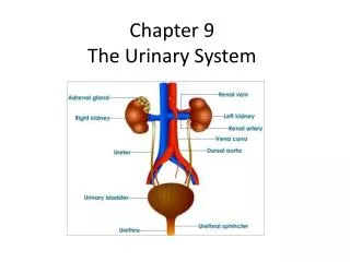
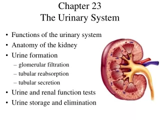
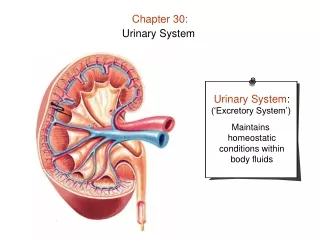
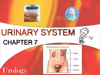
Components ---kidney : produce urine ---ureters ---bladder -cavity organs, conduct, store and ---urethra remove the urine ---functions: • remove waste products of metabolism • regulate the homeostasis • secrete some bioactive factors- renin, erythropoietin
*cavity organs: • mucosa: /epi-transitional epi /lamina propria • muscularis: SM • adventitia: CT
2. Kidney ---bean-shaped, 10-12cm in length, 5-6cm in width, 3-4cm in thickness ---hilum: BV, LV, N and ureters enter or out *renal pelvis: funnel-shaped expension of upper end of ureter *calyx: branches of renal pelvis
1)General structure: ---capsule: DCT ---cortex: dark stained /medullary ray /cortical labyrinth ---medulla: slight stained /renal pyramid /renal columns /renal papillae: -minor calyx -major calyx -pelvis
*renal lobe: one renal pyramid and its bounding cortical tissue *renal lobule: renal tissue including one medullary ray and cortical tissue surround it
According to function, renal parenchyma is mainly consists of uriniferous tubules ---parenchyma: /uriniferous tubules= renaltubule+ collecting tubules /renal corpuscle =glomerulus + renal capsule(beginning part of renal tubule) /nephron=renal corpuscle + renal tubule ---interstitium: CT, BV, N
2)Nephron: structural and functional unit, 1,000,000 ---renal corpuscle: cortical and juxtamedullary nephron /renal glomerulus: /renal capsule ---renal tubule: /proximal tubule: -convoluted portion -straight portion /thin segment /distal tubule: -straight portion -convoluted portion
①renal corpuscle: spherical, 200um in D, two poles: vascular pole and urinary pole ---glomerulus: afferent arterioles→capillary network →efferent arterioles *capillary network: /endothelial cell: pore, 70-90 nm, no diaphragm /basal lamina: 330nm
*intraglomerular mesangium: /mesangial cell: -small, irregular, with processes -small dark N -EM: RER, Golgi, lysosome, phagocytic vesicles, cytoskeleton-MF, MT,IF and secretory granules -functions: i. produce ground substance ii. phagocytosis iii.contract iv.secrete renin and enzymes /matrix: -type IV collagen -glycoprotein: sulphate chondritin, heparin and dermatan
---renal capsule: two layers capsule –formed by beginning part of renal tubule which is enlarged and invaginated /parietal layer: simple squamous epi. /visceral layer: podocytes /renel capsular cavity: the space between two layers
*podocyte: • cells with many processes ( primary and secondary processes-foot processes) • Foot processes: interdigitated with each other and embraced the capillaries, • slit pore: -narrow intercellular space between foot processes -25 nm, with 4-6 nm diaphragm-slit membrane
* filtration barrier or membrane: the structure for filtration is called filtration barrier or membrane, including: • fenestrated endothelial cell: negative ions • basal lamina: type IV collagen, proteoglycan, laminin-negative ions (sulphate heparin) • slit membrane-nephrin(size selective filter: negative ions
②renal tubule: a. proximal tubule: 50-60um in D, 14 mm long ---structure LM: EM: • pyramidal cuboidal • eosinophilic • round N • brush-liked border - microvilli(apical canaliculi) • longitudinal striation- plasma membrane infolding • no clear boundary - lateral extension (rich in Na+ K+ ATPase)
---Function: i. reabsorption : -85% Na+ ions and water -All of glucose, aminoacid, polypeptide, proteins and vitamin -50% bicorbonated salt and sulphate salt ii. secrete H+, NH3, hippuric acid and creatinine
b. thin segment: /10-15 um, simple squamous epi, /facilitate the passage of water and ions /reabsorb Na+ and Cl- and 5% water
c. distal tubule: ---structure: LM: EM: • cuboidal • slight-stained • round N • no brush-liked border – less microvilli • well-developed longitudinal striation – plasma membrane infolding
Distal tubule Plasma membrane infolding
---function: i.reabsorption of 8% water, Na+ ions ii.excretion of K+, H+,NH3 iii. regulated by aldosterone(adrenal gland) and antidiuretic hormone (vasopressin) (pituitary gland)
3)collecting tubule: • arched collecting tubules • cortical collection tubules • medullary collection tubules -simple cuboidal epi. to simple columnar epi. -slight –stained -have clear boundary -reabsorb 4% water
a. juxtaglomerular cell: ---a groups of modified SM cell of afferent arterioles ---structure: -larger, cuboidal in shaped, with round N -contain secretory granules ---function: i.secrete renin→adrenal gland→aldosterone→blood pressure↑ ↑ angiotensinogen→angiotensin I→angiotensin II→contraction of SM of BV ii.secrete erythropoietin to promote erythropoiesis
b. macula densa ---a group of cells derived from epi. of distal tubule ---cell becomes taller, narrow, with round N apical part arranged ---cells have processes connecting with other cells ---function: chemoreceptors- feel the Na+ ions concentration
c. extraglomerular mesangial cell(polar cushion cell) ---similar to intraglomerular mesangial cell ---transfer the information d. peripolar cell ---structure: EM: -microvilli -junctional complexes -RER, Golgi, and granules ---function: regulate the reabsorption and secretion of renal tubule
5) renal interstitial: CT ---fibers: type I,III,IV collagen ---matrix ---cell: • fibroblast • macrophage • lipid-laden interstitial cell: -stellate cell with processes -osmiophilic lipid droplets: -function: i.involve in formation of F and matrix ii. secret prostaglandin
6) Blood supply to the kidney i. Very large blood flow (1.2L/min) ii. To form cap.two times iii. Glomerular cap. have a high blood pressure iv. Form vasa recta loop near medullary loop v. More larger blood flow in renal cortex
Interlobar arteries → Arcuate arteries →Interlobular arteries →Capsular cap. ↑ ↓ ↓ Renal artery Afferent arterioles Afferent arterioles ↓ ↓ Juxtamedullary nephron Cortical nephron glomerulus glomerulus ↓ ↓ Efferent arterioles Efferent arterioles ↓ ↓ Vasa recta(artery and vein)← Capillary network Capillary network ↓ ↓ ↓ Renal vein←Interlobar vein← Arcuate vein ← Interlobular vein ← Stellate vein