
Uploaded by
ira
8 SLIDES
233 VUES
80LIKES
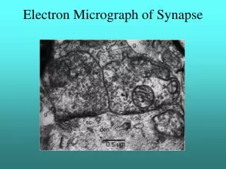
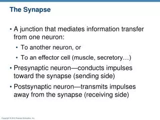
Electron Micrograph of Synapse
DESCRIPTION
This electron micrograph provides a detailed view of synaptic structures within the brain, specifically focusing on the brainstem, limbic system, and various lobes of the cerebral cortex. The image highlights key anatomical features including the thalamus, lateral geniculate nucleus (LGN), striate cortex (V1), and the organization of parvocellular and magnocellular layers. These insights are crucial for understanding neural connectivity and information processing within visual pathways.
Download

1 / 8
Télécharger la présentation 
Electron Micrograph of Synapse
An Image/Link below is provided (as is) to download presentation
Download Policy: Content on the Website is provided to you AS IS for your information and personal use and may not be sold / licensed / shared on other websites without getting consent from its author.
Content is provided to you AS IS for your information and personal use only.
Download presentation by click this link.
While downloading, if for some reason you are not able to download a presentation, the publisher may have deleted the file from their server.
During download, if you can't get a presentation, the file might be deleted by the publisher.
E N D
Presentation Transcript
LGN to V1 Striate Cortex or LGN V1 Parvocellular Layers Interblobs Blobs Layer 4b Magnocellular Layers


More Related