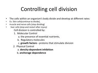
Controlling cell division
Controlling cell division. The cells within an organism’s body divide and develop at different rates: Ex. Skin cells(continue to divide), muscle and nerve cells (stop dividing) liver cells (stop and restart after injury) Cell division is controlled by: 1. Molecular Control

Controlling cell division
E N D
Presentation Transcript
Controlling cell division • The cells within an organism’s body divide and develop at different rates: • Ex. Skin cells(continue to divide), • muscle and nerve cells (stop dividing) • liver cells (stop and restart after injury) Cell division is controlled by: 1. Molecular Control a. the presence of essential nutrients, b. Regulatory molecules c. growth factors - proteins that stimulate division 2. Physical Control a. density-dependent inhibition b. anchorage dependence
Molecular Control Systempresence of certain chemicals triggers next phase • There are three major checkpoints in the cell cycle. (G1, G2, M) • G1 checkpoint • a. allows entry into the S phase or b. causes the cell to leave the cycle, entering a nondividing G0 phase 2. G2 checkpoint 3. M - mitosis
G0 G1 S G2 M checkpoint G2 checkpoint
1. G1 – Restriction Point • 1. most important • A. If receives go ahead will divide • B. No go ahead –> G0 (non-dividing state) Involves Chemical triggers i.e. Growth Factors – - proteins secreted by body cells that trigger cells to divide Ex. PDGF Most work at G1 checkpoint through a Signal Transduction Pathway
Figure 8.8B EXTRACELLULAR FLUID Plasma membrane Growth factor Relay proteins G1 checkpoint Receptor protein Signal transduction pathway S G1 Control system M G2 CYTOPLASM
Cultured cells suspended in liquid The addition of growth factor
G2 Checkpoint • Involves Regulatory molecules (chemical triggers) • A. proteins involved – kinases and cyclins • Kinases - Enzymes that phosphorylate other proteins (activate or inactivate) • Cyclins – proteins that fluctuate in cell • Cyclin + kinases • cyclin-dependent kinases (cdks)
Figure 8.8A As cell grows (G1, S, G2) cyclin levels increase At G2 –high level of cyclins Cdks form and become mitosis promoting factors (MPF) MPF fragments nuclear envelope (phosphorylation) MPF destroys cyclin – levels Cyclins increase G1 checkpoint G0 S G1 M G2 G2 checkpoint
Physical Control Anchorage Density dependent inhibition crowded cells stop dividing Cells reach high density have insufficient growth factors and nutrients Single layer of cells 2. Anchorage dependence Removal of cells a. Cells must be in contact with a solid surface Restoration of single layer by cell division
Cancer • Abnormal cells: • 1. Do not exhibit: • -density –dependent inhibition • - anchorage dependence • 2. Cell undergoes transformation • - tumor results - 2 types • Benign/ malignant