Tissue Repair
Tissue Repair. Dr. Raid Jastania. What is Repair? When does regeneration occur? When does fibrosis occur? What are the consequences of fibrosis?. Intended Learning Outcomes: Student should understand the relation between inflammation and repair process.

Tissue Repair
E N D
Presentation Transcript
Tissue Repair Dr. Raid Jastania
What is Repair? • When does regeneration occur? • When does fibrosis occur? • What are the consequences of fibrosis?
Intended Learning Outcomes: • Student should understand the relation between inflammation and repair process. • Student should be able to discuss the types of repair (regeneration and fibrosis). • Student should know how regeneration occurs • Students should know how scarring and fibrosis occurs • Student should be able to apply the concept of inflammation and repair to describe the pathogenesis of chronic inflammatory diseases. eg. Rheumatoid arthritis, Liver cirrhosis, and pulmonary fibrosis.
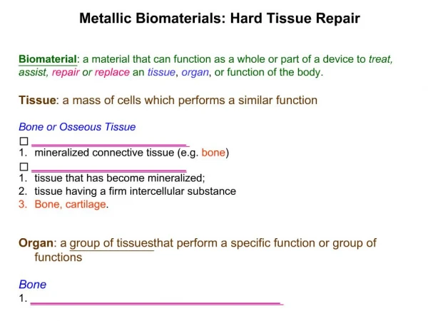
Repair • It is the response of tissue to restore the normal structure and function following injury. Repair process starts early with inflammation and it consists of: • Regeneration of parenchymal cells • Replacement of injured tissue by connective tissue (fibrosis)
Regeneration of Parenchymal cells • In normal adults, mature cells have the ability to keep the same number of cells in certain tissues and organs. • This results from the balance between cell proliferation on one side, and cell death/apoptosis or differentiation on the other.
The cell cycle • These phases are: • G1: Pre synthetic phase • S: Synthetic phase • G2: Pre mitotic phase • M: mitosis • G0: Not dividing cells. Cells not in the “cycle”
The cell cycle • Cyclins and CDK’s (cyclin-dependent kinases). • For example: Cyclin B level increases during G2. Cyclin B activates CDK1. Following mitosis both Cyclin B and CDK1 are degraded by ubiquitin-proteasome pathway.
The cell cycle • CDK’s are also regulated by CDK inhibitors. • Example of CDK inhibitor is P53. P53 acts as a break in the cell cycle at the G1-S “Check point” It allows repair of DNA damage during replication. This happens by enhancing the expression of P21 (CDKNIA) which arrest the cycle in G1. If DNA repair occurs, P53 let the cell pass to S phase. However if DNA repair is not possible, P53 forces the cell to die by apoptosis.
Proliferation Ability • Labile cells. These are the cells that continuously divide. These cells have the ability to renew the stem cells “self-renewal” and to differentiate. • Stable cells. These are the cells that can divide and proliferate only when they are stimulated (usually by injury). This is like parenchymal cells of liver, kidney, pancreas, and endothelium, fibroblast and smooth muscle cells. • Permanent cells. These are terminally differentiated cells that cannot divide. This includes neurons and cardiac muscle cells.
Fibrosis (Repair by connective tissue) • This occurs when the injury is severe resulting in parenchymal cell death and loss of the normal stromal framework.
Fibrosis (Repair by connective tissue) • Components: • Angiogenesis • Fibrosis Migration and proliferation of fibroblasts Deposition of extracellular matrix (ECM) 3. Maturation and reorganization (Remodeling)
Angiogenesis neovascularization Steps: • Proteolytic degradation of parent vessel basement membrane (BM) • Migration of endothelial cells toward angiogenic stimulus • Proliferation of endothelial cells • Maturation of endothelial cells and organization of capillary tubes by formation of pericytes, and smooth muscle cells..
Angiogenesis neovascularization Angiogenesis ends by formation of “Granulation tissue”: Granulation tissue is pink granular tissue seen at site of tissue repair. It is formed by new vessels and loose connective tissue, often mixed with inflammatory cells.
Angiogenesis neovascularization Factors induce angiogenesis: • bFGF (basic fibroblast growth factor), VEGF (vascular endothelial growth factor) • Produced by many stromal cells • Action: stimulate endothelial cell proliferation and production of proteinases
Fibrosis • Fibrosis is built on granulation tissue. • Steps: • Emigration and Proliferation of fibroblasts • Stimulated by PDGF (platelet derived growth factor), bFGF, TGF-beta (transforming growth factor beta) • Produced by endothelial cells and inflammatory cells • Deposition of ECM: • Collagen synthesis is stimulated by PDGF, bFGF, TGF-beta, TNF, IL-1
Scar Remodeling • The transition from granulation tissue to scar. • Remodeling result from the net balance between ECM synthesis and degradation. • Metalloproteinases are produced by fibroblasts, macrophages, neurophils, synovial cells to degrade ECM. • The process is controlled by GF’s, cytokines… and inhibited by TGF-beta
Extracellular Matrix (ECM) • There are 2 forms of ECM: • Interstitial matrix: collagen, proteoglycans, and glycoproteins • Basement membrane • Function: Mechanichal support Cell orientation (polarity) Control of growth Maintenance of differentiation Scaffolding for tissue renewal Storage of regulatory molecules
Extracellular Matrix (ECM) • Components: • Structural proteins • Gel • Adhesive glycoproteins
Collagen More than 30 peptide chains and 18 types of collagen. Collagen type I in bone, type II in cartilage, type III in reticulin framework, type IV in basement membrane It provides the tensile strength to tissues.
Elastin: • Provides elasticity. Found in vessels, skin, ligaments • Proteoglycans and hyaluronan: • It forms the extracellular gel. • Proteoglycans are long polysaccharides (glycosaminoglycans)
Adhesive glycoproteins and integrins: • Fibronectin: synthesized by fibroblasts, monocytes, and endothelium • Fibronectin binds to collagen, fibrin, proteoglycans • Laminin found in basement membranes • Integrins bind to ECM