Download
1 / 64
730 likes | 1.29k Vues
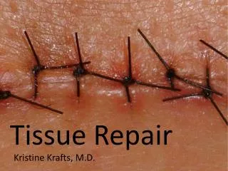
Tissue Repair. Kristine Krafts, M.D. Tissue Repair. Tissue repair = restoration of tissue architecture and function after an injury Occurs in two ways: Regeneration of injured tissue Replacement by connective tissue (scarring) Usually, tissue repair involves both processes

E N D
Tissue Repair Kristine Krafts, M.D.
Tissue Repair • Tissue repair = restoration of tissue architecture and function after an injury • Occurs in two ways: • Regeneration of injured tissue • Replacement by connective tissue (scarring) • Usually, tissue repair involves both processes • Involves cell proliferation, and interaction between cells and extracellular matrix
Tissue Repair Lecture Outline • Important background facts • Cellular proliferation • Growth factors • The extracellular matrix • The process of tissue repair • Regeneration • Scarring • An illustration: skin wound healing • Why do good wounds go bad?
Tissue Repair Lecture Outline • Important background facts • Cellular proliferation
Cellular Proliferation • Lots of cells proliferate during tissue repair: • Injured tissue remnants • Vascular endothelial cells • Fibroblasts • You need to know a few things about: • The cell cycle • The proliferative capacities of different tissues • Stem cells • Growth factors • The extracellular matrix
Cellular Proliferation Tissues of the body are divided into three groups: • Continuously dividing (labile) tissues • Stable tissues • Permanent tissues
Cellular Proliferation Tissues of the body are divided into three groups: • Continuously dividing (labile) tissues • Cells are continuously proliferating • Can easily regenerate after injury • Contain a pool of stem cells • Examples: bone marrow, skin, GI epithelium
Cellular Proliferation Tissues of the body are divided into three groups: • Continuously dividing (labile) tissues • Stable tissues • Cells have limited ability to proliferate • Limited ability to regenerate (except liver!) • Normally in G0, but can proliferate if injured • Examples: liver, kidney, pancreas
Cellular Proliferation Tissues of the body are divided into three groups: • Continuously dividing (labile) tissues • Stable tissues • Permanent tissues • Cells can’t proliferate • Can’t regenerate (so injury always leads to scar) • Examples: neurons, cardiac muscle
Tissue Repair Lecture Outline • Important background facts • Cellular proliferation • Growth factors
Growth Factors • Very important in tissue repair. • Actions: • Stimulate cell division and proliferation • Promote cell survival • Huge list! Usually have “GF” in name: • EGF (epidermal growth factor) • TGF (transforming growth factor) • PDGF (platelet-derived growth factor)
Tissue Repair Lecture Outline • Important background facts • Cellular proliferation • Growth factors • The extracellular matrix
The Extracellular Matrix • ECM is the network that surrounds cells • Two forms: interstitial matrix and basement membrane • Does lots of things! • Sequesters water and minerals • Gives cells a scaffold to adhere to • Stores growth factors
Extracellular Matrix • Bottom line: ECM regulates proliferation, movement, and differentiation of the cells living in it. • If you screw up your ECM, you can’t regenerate! You’ll form a scar instead.
Tissue Repair Lecture Outline • Important background facts • Cellular proliferation • Growth factors • The extracellular matrix • The process of tissue repair • Regeneration
Regeneration • Occurs all the time in labile tissues • Cells are constantly being lost and replaced • If demand increases, supply increases easily • Occurs in limited form in stable tissues • Remove one kidney: the other one undergoes hypertrophy and hyperplasia • Remove half of the liver: it will grow back • Only occurs if residual tissue is intact!
Liver before resection right lobe to be resected left lobe now enlarged Liver 1 week after resection
Tissue Repair Lecture Outline • Important background facts • Cellular proliferation • Growth factors • The extracellular matrix • The process of tissue repair • Regeneration • Scarring
Scarring • If injury is severe, regeneration can’t happen • So, fibrosis (a scar) replaces the injured tissue • Four components to this process: • New vessel formation (angiogenesis) • Fibroblast proliferation • Synthesis of collagen (scar formation) • Remodeling of scar
Scarring • By 24 hours: • Endothelial cells start proliferating • Fibroblasts emigrate • By 3-5 days: • Granulation tissue present • Weeks later: • Dense fibrosis (scar) • Scar is remodeled over time
Scarring Summary: 1. Make granulation tissue 2. Turn it into a chunk of collagen
Q. Is “granulation tissue” the same thing as“granuloma”?
Q. Is “granulation tissue” the same thing as“granuloma”? A. No!
Q. Is “granulation tissue” the same thing as“granuloma”? A. No! Q. Is it the same thing as “chronic granulomatous disease”?
Q. Is “granulation tissue” the same thing as“granuloma”? A. No! Q. Is it the same thing as “chronic granulomatous disease”? A. No!
Tissue Repair Lecture Outline • Important background facts • Cellular proliferation • Growth factors • The extracellular matrix • The process of tissue repair • Regeneration • Scarring • An illustration: skin wound healing
Skin Wound Healing • First intention • Second intention
first intention healing second intention healing
Healing by First Intention • Occurs in small wounds that close easily • Epithelial regeneration predominates over fibrosis • Healing is fast, with minimal scarring/infection • Examples: • Paper cuts • Well-approximated surgical incisions • Replaced periodontal flaps
Healing by First Intention • By 24 hours • By 3-7 days • Weeks later
Healing by First Intention • By 24 hours • clot forms • neutrophils come in • epithelium begins to regenerate
Healing by First Intention • By 24 hours • By 3-7 days • Macrophages arrive • Granulation tissue is formed • Collagen begins to bridge incision • Epithelium increases in thickness
Healing by First Intention • By 24 hours • By 3-7 days • Weeks later • Granulation tissue is gone • Collagen is remodeled • Epidermis is full and mature(but without dermal appendages) • Eventually, scar forms
24 hours 6 hours 2 days 1 week
Healing by Second Intention • Occurs in larger wounds that have gaps between wound margins • Fibrosis predominates over epithelial regeneration • Healing is slower, with more inflammation and granulation tissue formation, and more scarring. • Examples: • Infarction • Large burns and ulcers • Extraction sockets • External-bevel gingivectomies
Skin Wound Healing • More inflammation • More granulation tissue • Wound contraction Second intention healing has: