Burns in Children Review
810 likes | 1.85k Vues
Burns in Children Review. Tarek Hazwani, MD Assistant Consultant Pediatric Intensivist King Abdulaziz Medical City. Burns in Children Review. Anatomy of Skin Pathophysiology Critical Factors Management. Anatomy of Skin. Largest body organ More than just a passive covering. Anatomy.

Burns in Children Review
E N D
Presentation Transcript
Burns in Children Review Tarek Hazwani, MD Assistant Consultant Pediatric Intensivist King Abdulaziz Medical City
Burns in Children Review • Anatomy of Skin • Pathophysiology • Critical Factors • Management
Anatomy of Skin • Largest body organ • More than just a passive covering
Anatomy • Two layers • Epidermis • Dermis
Skin Functions • Sensation • Protection • Temperature regulation • Fluid retention
Burn: Pathophysiology • Loss of fluids • Inability to maintain body temperature • Infection
Burn: Pathophysiology • Patients with large burns (≥15 percent TBSA for young children and ≥20 percent for older children and adolescents) develop systemic responses to these mediators. • For patients with 40 percent TBSA or more, myocardial depression can occur . • As a result, patients with major burns may become hypotensive (burn shock) and edematous (burn edema).
Burn: PathophysiologyMetabolic response : • Following resuscitation, children with major burns develop a hypermetabolic response that results in a dramatic increase in energy expenditure and protein metabolism . • Evidence suggests that modulation of the hypermetabolic response with therapies such as beta blockers and human growth hormone may improve outcomes for severely burned children
Pathophysiology • Systemic capillary leak usually persists for 18 to 24 hours. Protein is lost from the intravascular space during the first 12 to 18 hours after a burn, after which vascular integrity improves.
Critical Factors • Depth • Extent
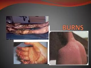
Burn Depth • First Degree (Superficial) • Involves only epidermis • Red • Painful • Tender • Blanches under pressure • Possible swelling, no blisters • Heal in ~7 days
Burn Depth • Second Degree (Partial Thickness) • Extends through epidermis into dermis • Salmon pink • Moist, shiny • Painful • Blisters may be present • Heal in ~7 to 21 days
Burn Depth • Burns that blister are second degree. • But all second degree burns don’t blister.
Burn Depth • Third Degree (Full Thickness) • Through epidermis, dermis into underlying structures • Thick, dry • Pearly gray or charred black • May bleed from vessel damage • Painless • Require grafting
Burn Depth • Often cannot be accurately determined in acute stage • Infection may convert to higher degree • When in doubt, over-estimate
18 9 9 18, Front 18, Back 1 13.5 13.5 Burn Extent • Pediatric Rule of Nines • For each year over 1 year of age, subtract 1% from head, • add equally to legs.
Burn Extent • Rule of Palm • Patient’s palm equals 1% of his body surface area
Burn Severity • Based on • Depth • Extent • Location • Cause • Patient Age • Associated Factors
Critical Burns Need Burn centreAmerican Burn Association • Age <10 years with >10 percent TBSA burn • Age ≥10 years with >20 percent TBSA burn • Full thickness burn >5 percent TBSA • Inhalational injury • Any significant burn to face, eyes, ears, genitalia, or joints • Significant associated injuries (fractures or major trauma)
Associated Factors • Patient Age • < 5 years old • > 55 years old • Burn Location • Circumferential burns of chest, extremities
Burn shock • characterized by specific hemodynamic changes (decreased cardiac output and plasma volume, increased extracellular fluid, and oliguria)
Burn Edema • Fluid shift intravascular to extravascular soon after a burn—persist for the first 24 hours • In small burns edema peaks early, in large burns edema developed continue for 18-24 hours • Unburned tissue edema occurs when burn exceeds 35-40% TBSA • Early increase vascular permeability—in part related to histamine—mechanism is likely related to PMN and their adhesion to the endothelium
Stop Burning Process • Remove patient from source of injury • Remove clothing unless stuck to burn • Cut around clothing stuck to burn, leave in place
Assess Airway/Breathing • Start oxygen if: • Moderate or critical burn • Decreased level of consciousness • Signs of respiratory involvement • Burn occurred in closed space • History of CO or smoke exposure • Assist ventilations as needed
Assess Circulation • Check for shock signs /symptoms Early shock seldom results from effects of burn itself. Early shock = Another injury until proven otherwise
Obtain History • How long ago? • What has been done? • What caused burn? • Burned in closed space? • Loss of consciousness? • Allergies/medications? • Past medical history?
Rapid Physical Exam • Check for other injuries • Rapidly estimate burned, unburned areas • Remove constricting bands
Treat Burn Wound • Cover with DRY, CLEAN SHEETS • Do NOT rupture blisters • Do NOT put goo on burn
Special Considerations • In Pediatrics always Consider possibility of abuse • As many as 10% of abuse cases involve burns
Burn: Management • Parkland formula, as follows: • (2-4 cm3 of crystalloid) X (% BSA burn) X (body weight in kg) • The Parkland formula must be modified in pediatric patients by adding maintenance
Burn: Management Fluid resuscitation • Estimating fluid requirements : for the first 24 hours following a burn injury include: • Parkland - 4 mL/kg per percent total burn surface area (TBSA). Add glucose maintenance fluid for children <5 years of age. • Galveston - 5000 mL/m2 per percent TBSA. Add 2000 mL/m2 per day for maintenance requirements. • Half of the fluid is given over the first 8 hours. The remaining half is given over the next 16 hours
Burn: Management Fluid resuscitation Choice of fluid: • Ringers lactate (RL) is the resuscitation and maintenance fluid of choice for the first 24 hours at most burn centers. • Experts recommend adding D5% to maintenance fluid for children <20 kg to prevent hypoglycemia . • Colloid is typically added after 24 hours to restore oncotic pressure and preserve intravascular volume
Burn: Management Colloid resuscitation • The addition of plasma or albumin to resuscitation fluids has been criticized on the assumption that the burn-induced increase in vascular permeability and the consequent extravasation of proteins persist for up to 36 h post injury . • The main concern is that protein administration during the first 24 h increases protein accumulation in the interstitium and thus traps water . • Using 131iodine-labeled albumin and autoradiographic techniques to demonstrate have shown that effective transcapillary sieving of albumin molecules into burned skin essentially stops at approximately 8 h post injury and that edema of injured tissues, maximal at 3 h post burn, persists beyond24 h post injury
Burn: Management Fluid resuscitation Monitoring fluid status: • The volume status of burn patients must be carefully monitored in order to successfully navigate the narrow path between inadequate volume and fluid overload. The following parameters are helpful : • Urine output should be maintained at 1 to 2 mL/kg per hour for children <30 kg and 0.5 to 1 mL/kg per hour for those ≥30 kg. • Heart rate is a better monitor of circulatory status in children than is blood pressure. Tachycardia may indicate hypovolemia, but pain can elevate heart rate in euvolemic patients. • Metabolic acidosis can be a marker for inadequate fluid resuscitation, but also occurs with carbon monoxide or cyanide exposure
Burn: Management Fluid resuscitation Burn Children not response to large fluid volumes to maintain adequate perfusion : • Volume loss from occult injuries • Neurogenic shock as the result of a spinal cord injury • Myocardial depression or decreased vascular tone from inhaled or ingested toxins
Burn: Management Pain control • Most burn centers use • morphine • Fentanyl may be a safer choice for initial pain management for patients whose cardiovascular status may be unstable
Burn: Management Antibiotics • Topical antibiotics have been used to dress burn wounds: • It is : available, and reduce the risk of infection. • The topical antibiotic is applied to the wound which is then covered with a nonadherent dressing. • Specific antibiotic : Silver sulfadiazine , Mafenide , Bacitracin
Burn: ManagementSpecial Considerations • Steroids have no role in treating burn wounds • Intravenous antibiotics are not recommended in the initial treatment of most burn patients, as it may increase the chance of colonization with more virulent and resistant organisms. They should be reserved for those patients with secondary infections
Burn: ComplicationsInfection • Early Infections : • Organism : GAS , S. aureus • Specific colonization of burn wounds is somewhat predictable over time. Initially, gram-positive organisms are present; • infection that occurs in the first 48 hours after the burn is usually secondary to GAS. • The incidence of GAS infections in burned patients has decreased, probably secondary to immediate use of topical antimicrobial therapy. • Routine administration of antibiotics prophylaxis is not recommended ( colonization and potential infection with more resistant organisms). • S. aureus also causes early septicemia. If there is concomitant inhalation injury.
Burn: ComplicationsBacteremia • Bacteremia is not uncommon in the burned patient. • Risk factors include wound manipulation and the presence of an intravascular catheter. • infected intravascular thrombus can cause persistent bacteremia. • Endocarditis must be considered in any patient with prolonged bacteremia.
Burn: ComplicationsRenal failure • ARF in burn patients is not common. Two distinct pictures • of ARF can be observed: early ARF, occurring • either few hours after injury or in the first few days, • and late ARF developing approximately 1 or more • weeks after burn injury. Early ARF may be due to • hypovolemia and hypoperfusion of the kidneys, • whereas late ARF is a consequence of infection, endotoxemia, • and MODS
Burn: ComplicationsRenal failure • Renal damage can arise even from hemoglobinuria • in burn patients with associated hemolysis, the administration of haptoglobin may prevent hemoglobinuria-nduced renal failure
Inhalation Injury • 10-20% hospitalized burn patients sustained inhalation injury. • Increased mortality • History (closed space) • P.E. (facial burn, singed nasal hairs, erythema, carbonaceous material in back of the troat) • laboratory tests (carboxyhemoglobin>15%) and bronchoscopy (erythema and sooty deposite in the airway) • Treatment: supportive. Nasotracheal or endotracheal intubation preferable to early tracheostomy. Prophylactic antibiotics and steroids not indicated.
Inhalation Injury Problems • Hypoxia • Carbon monoxide toxicity • Upper airway burn • Lower airway burn
Inhalation Injury Carbon Monoxide • Product of incomplete combustion • Colorless, odorless, tasteless • Binds to hemoglobin 200x stronger than oxygen • Headache, nausea, vomiting, “roaring” in ears
Inhalation Injury Carbon Monoxide Exposure makes pulse oximeter data meaningless!
Inhalation Injury Carbon Monoxide Measurement • Carbon monoxide has various effects depending upon levels • Must check levels on Blood Gas analysis 0-10% can be seen in smokers can be seen in smokers 10-20% patients can have headache 20-30% patients develop severe headache, nausea, vomiting, CNS collapse 30-40% patients present with syncope, convulsions, depressed cardiac activity and respiratory function 40% and greater death may ensue within hours