Burns
Burns. DR BABAK MASOUMI ASSISTANT PROFESSOR OF EMERGENCY MEDICINE ESFAHAN UNIVERSITY of MEIDCAL SCIENCES 2011. Classification of Burns. Superficial Superficial partial-thickness Deep partial-thickness Full-thickness. Superficial.

Burns
E N D
Presentation Transcript
Burns DR BABAK MASOUMIASSISTANT PROFESSOR OF EMERGENCY MEDICINEESFAHAN UNIVERSITYof MEIDCAL SCIENCES 2011
Classification of Burns Superficial Superficial partial-thickness Deep partial-thickness Full-thickness
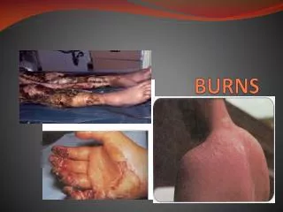
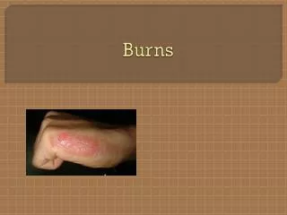
Superficial Very painful, dry, red burns which blanch with pressure. They usually take 3 to 7 days to heal without scarring. Also known as first-degree burns. The most common type of first-degree burn is sunburn.First-degree burns are limited to the epidermis, or upper layers of skin.
Superficial Partial-Thickness Very painful burns sensitive to temperature change and air exposure. More commonly referred to as second-degree burns. Typically, they blister and are moist, red, weeping burns which blanch with pressure. They heal in 7 to 21 days. Scarring is usually confined to changes in skin pigment.
Deep Partial-Thickness Blistering or easily unroofed burns which are wet or waxy dry, and are painful to pressure. Their color may range from patchy, cheesy white to red, and they do not blanch with pressure. They take over 21 days to heal and scarring may be severe. It is sometimes difficult to differentiate these burns from full-thickness burns.
Full-Thickness Burns which cause the skin to be waxy white to a charred black and tend to be painless. Healing is very slow, if at all, and may require skin grafting. Severe scarring usually occurs.
The Skin The skin, the largest organ of the body, consists of two layers-the epidermis and dermis. The depth or degree of burn depends on which layers of skin are damaged or destroyed. The epidermis is the outer layer that forms the protective covering. The thicker or inner layer of the dermis contains blood vessels, hair follicles, nerve endings, sweat and sebaceous glands. When the dermis is destroyed, so are the nerve endings that allow a person to feel pain, temperature, and tactile sensation.
The most important function of the skin is to act as a barrier against infection. The skin prevents loss of body fluids, thus preventing dehydration. The skin also regulates the body temperature by controlling the amount of evaporation of fluids from the sweat glands. The skin serves a cosmetic effect by giving the body shape. When the skin is burned, these functions are impaired or lost completely. The severity of the skin injury depends upon the size of the injury, depth of the wound, part of the body injured, age of the patient, and past medical history. Because of the importance of the skin, it becomes clear that injury can be traumatic and life threatening. Recovery from burn injury involves four major aspects: burn wound management, physical therapy, nutrition, and emotional support.
The rule of nines • The rule of nines is a practical technique for estimating the extent of TBSA involved in a burn injury. • This approach divides the major anatomic areas of the body into percentages of TBSA. • For the adult, it allots 9% of the TBSA to the head and neck and to each upper extremity, 18% each to the anterior and posterior portions of the trunk, 18% to each lower extremity, and 1% to the perineum and genitalia. • The area of a patient's palm represents approximately 1% of the TBSA and can be helpful in calculating scattered areas of involvement.
The rule of nines • The primary difference between the TBSA of the adult and infant reflects the size of the infant's head (18%), which is proportionally larger than that of the adult ,and the lower extremities (14%), which are proportionately smaller than those of the adult.
Categorization of Patients • The severity of a burn injury depends on the (1) extent, depth. and location of the burn injury; (2) age of the patient; (3) etiologic agents involved; (4) presence of inhalation injury; (5) coexisting injuries or preexisting illnesses. • This classification creates three categories of burn injury (major, moderate. and minor) and defines the optimal setting for themanagement of each.
Major burn injury (1) partial-thickness burns involving more than 25% of TBSA in adults or 20% of TBSA in children younger than 10 and adults older than 50; (2) full-thickness burns involving more than 10% of TBSA; (3) burns involving the face, eyes. ears. hands. feet, or perineum that may result in functional or cosmetic impairment; (4) burns caused by caustic chemical agents; (5) high-voltage electrical injury; (6) burns complicated by inhalation injury or major trauma; and (7) burns sustained by high-risk patients (e.g.. those with underlying debilitating diseases).
Major burn injury • These injuries are best managed in a specialized burn center staffed by a team of professionals with expertise in the care of burn patients, including both acute care and rehabilitation.
Moderate burn injury includes (1) partial-thicknessof 15% to 25% of TBSA in adults or 10% to 20% of TBSA in children and older adults. (2) Fullthickness burns involving 2% to 10% of TBSA that do not present a serious threat of functional or cosmetic impairment of the eyes, ears. face, hands. feet, or perineum. The moderate category excludes high-voltage electrical injury. all burns complicated by inhalation injury or other trauma. and burns sustained by highrisk patients.
Moderate burn injury includes Patients with moderate burn injurie should be hospitalized for their initial care but not necessarily at a burn center.
Minor burn injury • partial-thickness burns involving less than 15% of TBSA in adults or 10% of TBSA in children and older adults. (2) fullthickness burns involving less than 2% of TBSA that do not present a serious threat of functional or cosmetic risk to eyes. ears. face. hands. feet. or perineum. These burns can usually be managed safely in the outpatient setting.
Treatment • Treatment should begin immediately to cool the area of the burn. This will help alleviate pain. • For deep partial-thickness burns or full- thickness burns, begin immediate plans to transport the victim to competent medical care. For any burn involving the face, hands, feet, or completely around an extremity, or deep burns; immediate medical care should be sought. Not all burns require immediate physician care but should be evaluated within 3-5 days. • Remove any hot or burned clothing.
Are you one of those people that stays up to date on the latest sports scores and plays? Improper use, handling, and storage of hazardous materials can lead to a different type of scoring… it’s called burn scoring which measures the percentage of the body burned. The score you rate on this chart can last you a lifetime.
Treatment 4.Use cool (54 degree F.) saline solution to cool the area for 15-30 minutes. Avoid ice or freezing the injured tissue. Be certain to maintain the victim’s body temperature while treating the burn. 5. Wash the area thoroughly with plain soap and water. Dry the area with a clean towel. Ruptured blisters should be removed, but the management of clean, intact blisters is controversial. You should not attempt to manage blisters but should seek competent medical help. 6. If immediate medical care is unavailable or unnecessary, antibiotic ointment may be applied after thorough cleaning and before the clean gauze dressing is applied.
Special considerations • Scalding-typically result from hot water, grease, oil or tar. Immersion scalds tend to be worse than spills, because the contact with the hot solution is longer. They tend to be deep and severe and should be evaluated by a physician. Cooking oil or tar (especially from the “mother pot”) tends to be full- thickness requiring prolonged medical care. • Remove the person from the heat source. • Remove any wet clothing which is retaining heat. • With tar burns, after cooling, the tar should be removed by repeated applications of petroleum ointment and dressing every 2 hours.
Looks and tastes great, right? You should see what a hot liquid will do to a child’s skin when the two come into contact. Be sure to keep hot liquids out of reach of small children.
Special considerations Flame a. Remove the person from the source of the heat. b. If clothes are burning, make the person lie down to keep smoke away from their face. c. Use water, blanket or roll the person on the ground to smother the flames. d. Once the burning has stopped, remove the clothing. e. Manage the persons airway, as anyone with a flame burn should be considered to have an inhalation injury.
Special considerations Electrical burns: are thermal injuries resulting from high intensity heat. The skin injury area may appear small, but the underlying tissue damage may be extensive. Additionally, there may be brain or heart damage or musculoskeletal injuries associated with the electrical injuries. a. Safely remove the person from the source of the electricity. Do not become a victim.
Special considerations b. Check their Airway, Breathing and Circulation and if necessary begin CPR using an AED (Automatic External Defibrillator) if available and EMS is not present. If the victim is breathing, place them on their side to prevent airway obstruction. c. Due to the possibility of vertebrae injury secondary to intense muscle contraction, you should use spinal injury precautions during resuscitation. d. Elevate legs to 45 degrees if possible. e. Keep the victim warm until EMS arrives.
Special considerations Chemical burns- Most often caused by strong acids or alkalis. Unlike thermal burns, they can cause progressive injury until the agent is inactivated. a. Flush the injured area with a copious amount of water while at the scene of the incident. Don’t delay or waste time looking for or using a neutralizing agent. These may in fact worsen the injury by producing heat or causing direct injury themselves.
Perform an ABCDEF primary survey • A—Airway with cervical spine control • B—Breathing • C—Circulation • D—Neurological disability • E—Exposure with environmental control • F—Fluid resuscitation
Airway Compromised or at risk of compromise? Intubate Yes Cause: Mechanical Escharotomies Carboxyhaemoglobin Intubate and ventilate Smoke inhalation Nebulisers Non-invasive ventilation Invasive ventilation Blast injury Invasive ventilation Chest drains No Breathing Compromised? Yes No Circulation Compromised perfusion to an extremity? Yes Escharotomies No Neurological Disability Impaired score on Glasgow coma scale? Consider: Hypoxia (carboxyhaemoglobin level?) Hypovolaemia Traumatic brain injury Yes No Exposure Fully assess burn area and depth Full examination for concomitant injuries Keep warm Fluids Calculate resuscitation formula based on surface area and time since burn
Initial burn management in ED • Assess burn size and depth • Establish good IV access and give fluids • Give analgesia • Place NG tube and Foley catheter • Take baseline blood samples • Dress wound • Perform secondary survey, reassess, and exclude or treat associated injuries • Arrange safe transfer to specialist burns facility
Airway Management • The natural history of upper airway burn injury is the development of edema that narrows the airway 12 to 24 hours after injury. • The tongue and oral mucosa become edematous much more quickly, within minutes to hours. • Consequently, intubation rather than observation is recommended in patients with signs of upper airway injury, such as stridor, inspiratory grunting.wheezing, or tachypnea.
Airway Management • Fiberoptic bronchoscopy is a simple. safe, and accurate method of diagnosing acute inhalation injury. • If the respiratory insufficiency is caused by a constricting eschar of the anterior thorax that limits respiratory excursion, escharotomy is imperative.
CO Poisening • CO is present in smoke, and its affinity for hemoglobin is 280 times that of oxygen. • A CO level is obtained for all patients with suspected inhalation injury. • Patients should receive 100% oxygen until their carboxyhemoglobin (COHb)level is less than 10% because the elimination half-life of COHb depends on oxygen tension.
CO Poisening • In room air, the half-life of CO-bound hemoglobinis 4 hours. • Under 100% oxygen, the half-life of CO-bound hemoglobin decreases to 45 minutes. • Administration of 100% oxygen increases the gradient for oxygen binding to hemoglobin, and unbound CO is exhaled through the lungs.
Cyanide Poisoning • Specific therapy for cyanide poisoning in patients with inhalation injury is another consideration. • Cyanide causes tissue hypoxia by uncoupling oxidative phosphorylation by binding to mitochondrial cytochrome. • Empirical treatment for cyanide toxicity should be considered for patients with unexplained severe metabolic acidosis associated with elevated central venous oxygen content, normal arterial oxygen content, and a low COHb level.
Circulation • Patients with moderate burns should have at least one large-bore IV line placed through unburned skin, and those with severe burns should have at least two lines initiated. • When a burn patient requires considerable fluid resuscitation or has evidence of cardiopulmonary disease, a central venous line is indicated.
Circulation • The microvascular injury caused by a burn produces increased vascular permeability with edema formation that results in ongoing plasma volume loss. • Maximal edema formation occurs 8 to 12 hours after injury for small burns and 24 hours after injury for large burns. • The purpose of fluid resuscitation is to restore effective plasma volume, avoid microvascular ischemia, and maintain vital organ function.
Circulation • The amount of fluid required varies with the patient's age, body weight, and extent of TBSA burned. • Typically, burns greater than 20% of TBSA require IV fluid resuscitation because the accompanying gastrointestinal ileus precludes sufficient oral intake.
Circulation • Although different formulas for fluid resuscitation have been recommended, all regimens emphasize that adequate resuscitation is evidenced by a normal urine output • 1 mL/kg/hr in children younger than 2 years, • 0.5 mL/kg/hr in older children, • At least 30 to 40 mL/hr in adults, • A normal sensorium, and stable vital signs.
Circulation • The Parkland formula for fluid resuscitation of burn patients is used as follows: LR solution (4 mL/kg/% TBSA burned) is administered intravenously in the first 24 hours, with one half given in the first 8 hours and the other half over the next 16 hours.
Circulation • The percent TBSA used for the formula includes only second- and third-degree burn injuries. • The fluid loss must be calculated from the time of injury. • Fluid replacement must take into account the fluid administered prehospital personnel.
Circulation • To avoid overhydration, patients with inhalation injuries should be resuscitated with substantially less than formula predictions, with acceptance of a urinary output in the range of 0.3 to 0.5 mUkg/hr.
Circulation • When endothelial integrity is restored 24 hours after the injury, some clinicians favor the administration of 5% albumin at 0.5 mL/kg/% TBSA to maintain dynamic forces between the extracellular spaces and the intravascular system. In addition, a low dose • Dopamine infusion (3 to 5 µg/kg/min) is beneficial in restoring renal and splanchnic blood flow in patients with major burn injury.
Circulation • Hypertonic saline resuscitation has been associated with an increased occurrence of acute tubular necrosis and hyperchloremic metabolic acidosis, which can exacerbate the metabolic acidosis of hypovolemic shock. • Therefore, hypertonic saline is not currently recommendedfor resuscitation of burn.
Analgesia • All patients with large burns should receive intravenous morphine at a dose appropriate to body weight. • The need for further doses should be assessed within 30 minutes.
Wound management • Estimating surface area and depth of a burn. • Washing the wound and removing any loose skin. • Deroofing the blisters for ease of dressing, except for palmar & Soles blisters. • Cover the wounds with sterile moist saline dressings. • If disposition is delayed,, cleanse with sterile saline,Apply topical antibacterial agent (e.g., silver sulfadiazine, bacitracin, or mafenide acetate). • Prophylactic antibiotics not indicated • Tetanus toxoid or immunoglobulin: 0.5 mL IM; 250 U IM once along with toxoid .
Criteria for referral to a burns centre • Associated airway injury • Partial thickness burns >5% of total body surface area in a child • Partial thickness burns >10% of total body surface area in an adult >1% full thickness burn • Partial or full thickness burns to face, perineum, external genitalia, feet and hands, and over joints • Circumferential injury • Chemical and electrical burns • Extremes of age • Intentional injury • Comorbidity
Key points • Perform a systematic assessment as with any trauma patient (don’t get distracted by the burn) • Beware of airway compromise • Provide adequate analgesia • Exclude any concomitant injuries • Discuss with a burns unit early • If in doubt, reassess