BURNS
BURNS. By PROF. MOHAMED A. EL GHARBAWI e -mail: elgharma2@yahoo.com Web site: www. dr-elgharbawi.com. OBJECTIVES. To know: D efinition & pathology of burns ( must know ) Assessment of depth & area of burns ( must know ) Treatment of burns ( must know ) Complications ( must know )

BURNS
E N D
Presentation Transcript
BURNS By PROF. MOHAMED A. EL GHARBAWI e-mail: elgharma2@yahoo.com Web site: www. dr-elgharbawi.com
OBJECTIVES To know: • Definition & pathology of burns (must know) • Assessment of depth & area of burns (mustknow) • Treatment of burns ( must know) • Complications ( must know) • Other types of burns (must know)
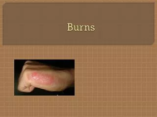
BURNS • DEFINITIONCoagulative necrosis of skin, mucous membranes and other tissues due to exposure to heat, electrical current, chemicals or irradiation.
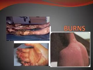
DEGREE OF BURN (DEPTH) • The deeper the burn, the longer the time for healing and more ugly and scaring outcome • 4 degrees of burns • SUPERFICIAL BURNS:Red in color and sensitive to pain stimulus (exposed nerve ends), include • First degree burnOnly erythema ( redness) of the skinMild burn with injury (no destruction) of the superficial layers of skin
DEGREE OF BURN (DEPTH) • Second degree burnBlebs & blisters are formedThere is some destruction of outer layers (epidermis) of skin • DEEP BURNSGray, white or black in color Not sensitive to pain stimuli (destroyed nerve ends)
DEGREE OF BURN (DEPTH) • Third degree Destruction of all layers of skin and skin appendages Epithelialization is not possible without skin grafts • Fourth degreeDestruction extends more to SC tissues, muscles or even to bones
EXTENT OF BURN (%) • It is the percentage of the total body surfacedamaged by the burn • Rule of 9 is used for estimationA. Adults: Head 9% Each upper limb 9% Each lower limb 18% (front 9% +back 9%) Front of trunk18% (Chest 9%+ Abdomen9%) Back of trunk 18% ( upper, mid , low back & buttocks) Genetalia 1%
EXTENT OF BURN (%) B. Children (modified) Head 18% Each upper limb 9% Anterior trunk 18% Posterior trunk 18% each lower limb 14% Genetalia 1%
EXTENT OF BURN (%) • Minor Burn 10% or less in children 15% or less in adults • Major Burn > 10% in children > 15% in adults • Mortality Minor burn rarely endanger life > 50% rarely survive • Burn of nasopharynx is dangerous , whatever extent
PATHOLOGY OF BURNS3 PHASES I. PHASE OF OEDEMA • First 3 -4 days • Massive inflammatory reaction+ Capillary injury • Hemorrhage into tissues + Thrombosis in blood vessels • Leak of fluids through the surface & into tissues leading to increased body weight • Fluid loss depends on extent & depth of burn
PATHOLOGY OF BURNSI.PHASE OF OEDEMA • 40% extent burn leads to severe &fatal fluid loss • 10% extent burn has minimal fluid loss • 3rd degree deep burn causes more fluid loss than other degrees • Very deep (4th degree) burn causes minimal fluid loss (destruction of all capillaries)
PATHOLOGY OF BURNSII.PHASE OF WOUND INFECTION • It takes about 30 days if the patient survives the edema phase • Sources of bacteria causing the infection1. From patient’s skin (found in crypts & hair follicles 2. Self contamination ( mouth, pharynx, anus) 3. From environment • Characterized by a –ve Nitrogen balance & weight loss • Weight loss due to loss of plasma from burn surface + increased tissue catabolism + sepsis
PATHOLOGY OF BURNSIII.PHASE OF HEALING &ANABOLSM • Next 30 days or more • If infection is controlled, the patient will be in good health & burn area is clean ( if it is grafted, it takes well) • If infection is renewed (Not controlled), burn area may get deeper & grafts melt away
CLINICAL PICTURE OF BURN • During Phase I In first few hours, patient seems not very ill, especially if you start treatment early & activelyPain is severe with superficial burns & less with deep destructive burns Patient is restless and thirsty Edema phase ends by diuresis & self correction of the electrolytes disturbance
CLINICAL PICTURE OF BURN • During Phase IINausea, Vomiting and may be GI ulceration ( 2nd part duodenum, Curling Ulcer)Anemia: Hemolysis + Loss from burn surface and GI ulcers + Malnutrition Loss of appetite Patient looks ill and depressed If infection is controlled, catabolism stops, Appetite improves, gradual gain of weight and burn starts to heal on 7th day If infection is not controlled, epithelium is destroyed and patient deteriorates
CLINICAL PICTURE OF BURN • During Phase IIIPatient’s appetite is increased Steady weight gainGrafts may look edematous, after few weeks lymphatic develop with improved muscle action leading to improve edema.
TREATMENT OF BURNS First Aid • Minimize contamination: Cover the burn by clean sheet, better soaked with iced water to minimize tissue destruction • Artificial respiration (mouth to mouth breathing) for respiratory arrest due to inhalation of steam
TREATMENT OF BURNS Treatment of minor burns • No fluid therapy is required • Cleaning • Apply antibiotic cream • Occlusive dressing (give rest to the burn area) • May be systemic antibiotic • Undress after 5 days to expose for dryness • Healing takes about 2 weeks
TREATMENT OF BURNS TREATMENT OF MAJOR BURNS A.General management during the first 48 hours 4. Tracheostomy may be needed in presence of respiratory irritation / obstruction (deep burns of face and neck) 5. Fluid therapy
TREATMENT OF BURNS TREATMENT OF MAJOR BURNS FLUID THERAPY • No fluids by mouth during the first 48 hours to avoid Gastric distension and paralytic ileus. • Given through 2 wide pore IV cannulas. • Better, through a central venous catheter (CV line) especially if IV hperalimentation is needed • Burns > 50% are considered as 50% when you calculate the needed fluid therapy.
TREATMENT OF BURNS TREATMENT OF MAJOR BURNS FLUID THERAPY Fluid therapy during the first 48 hours: Many formulas are used to calculate the amount of fluids. • BROOKE FORMULA • PARKLAND FORMULA
TREATMENT OF BURNS FLUID THERAPY BROOKE FORMULA First 24 hours: 1. Crystalloids (e.g. lactated Ringer’s solution) = 1.5 ml. X % of Burn surface area X Body weight in Kg 2. Colloids ( e.g. plasma od dextran) = 0.5 ml. X % of Burn surface area X body weight in Kg NB. Half of the calculated quantities are given over the first 8 hours & and second half is given over the next 16 hours. 3. Average daily water need for the adult is 2000 ml. to be given as glucose 5% (1000 ml. / 12 hours) Second 24 hours: Give half of the previously calculated crystalloids& colloids + Whole daily water need .
TREATMENT OF BURNS FLUID THERAPY PARKLAND FORMULA First 24 hours: 1. Crystalloids (lactated Ringer’s solution} 4 ml. X % of Burn surface area X Body weight in Kg 2. No Colloids 3. Daily water needs (2000 ml.) as glucose 5% Second 24 hours: Give half of the previously calculated amount of crystalloids + whole daily water need as glucose 5%
TREATMENT OF BURNS FLUID THERAPY PREAUTIONS Close observation & monitoring is mandatory with such groups of patients: 1. young children &old patients (Fragile) 2. Deep burns of head & neck (to avoid brain edema) 3. Patients with respiratory irritation ( to avoid pulmonary edema) 4. Patients with renal & cardiovascular diseases (to avoid overload)
TREATMENT OF BURNS EVALUATION OF FLUID THERAPY • Fluid deficiency is expected when the feel thirst, irritable and disoriented. Also, there are collapsed veins with hypotension. • Fluid excess is expected if there are crepitation over the lungs (pulmonary edema). Hypothermia and papilledema indicate brain edema. • Urine output of 0.5 ml. /kg/hour indicates a satisfactory fluid therapy. Fix a urinary catheter to measure the urine output during first 48 hours. • CVP measurement helps to adjust fluid therapy
TREATMENT OF BURNS B . General management after the first 48 hours • If Ht value < 40% , give more blood • If patient can't take his food for three days, give potassium with caution • If acute renal failure develops, give mannitol or low molecular weight dextran to improve the renal function
TREATMENT OF BURNS C. Management of burn wound • Aim is to control wound infection • Cleaning of burn wound under absolute aseptic conditions (sterile gowns& masks for all personnel attending the procedure) • Removal of all dead tissues from deep burns • Blisters & debris are removed under light GA or strong sedatives • Wound area is washed with sterile water/ saline with added antiseptic e.g. 1% Cetavlon • Burn coverage is the best method to prevent infection
TREATMENT OF BURNS DEFINITIVE LOCAL BURN CARE (3 methods) 1.Occlusive Dressing Method Covering the burn wound protects it against infection. Occlusive method is suitable for burns of extremities & burns affecting circumference of the trunk. Dressing should be: a. Occlusive (prevent infection) b. Absorptive (e.g. fine mesh gauze) to dry the burn area. c. Bulky and applied with even pressure ( to eliminate dead spaces & splint the area)
TREATMENT OF BURNS 2. Exposure Method • Exposure allows dryness of the burn wound with formation of crusts which works as cover to prevent contamination (mimic to closure). • Antibiotic powder to dust the burn until dryness • In superficial burn: exposure ends when crust fall. • In deep burn: exposure ends when healthygranulationtissue is formed and crusts is softened . Now, burn is ready for grafting after removal of crusts • Indication: Burns of Face, Perineum and One side of trunk
TREATMENT OF BURNS • TOPICAL ANTIMICROBIAL AGENTS a. Silver sulfadiazine cream: It is bactericidal for many G- ve and G+ve bacteria as wellas being effective against yeast. Used as local application for burns. b. Povidine -iodine ointment: Act against G- ve , G+ve and Fungi. Its use for long time in large burn areas may cause iodine toxicity
TREATMENT OF BURNS • BIOLOGICAL DRESSINGS • Porcine xenografts: available in the market.Daily application. Helps quick formation of healthy granulation tissue. • Homograft: difficult to obtain. Work as temporary life saving cover until rejection(2 – 4 weeks) • Human amnion and Biobrane (thin plastic membrane): Decrease fluid loss from burn
TREATMENT OF BURNS • HYDROTHERAPY It ranges from holding a small burn of a finger under the cold water to immersion of 50% burn patient in a hydrotank . It minimizes heat damage. Also, itRemoves dead tissue and pus. • Cleansthe surface of the wound . • Prevents loss of fluid through the skin • Protects the healthy tissue around the burn from trauma • Minimizes scar tissue formation • Minimize the risk of infection • Easy assessment of healing • Facilitates physiotherapy • Promotes formation of healthy tissue and healing
TREATMENT OF BURNS 3. EXCISION & GRAFTING • Immediate excision and grafting:For full thickness burns not > 15% surface area • Delayed excision and grafting:Crusts on deep burns treated by exposure can be excised or removed by repeated wet dressing or by use of proteolytic enzymes. Raw areas are covered by split thickness skin grafts • If donor sites are insufficient, use meshed skin graftsthat can expand up to 10 times. Such meshed grafts can’t be used on face, hands and flexor surfaces • Autogenous cultures of epithelium have been tried with some success
COMPLICATION OF BURNS • EARLY COMPLICATIONS:1. Neurogenic, Hypovolemic or Septic Shock2. Burn toxemia: Shock , Curling ulcer3. Infection: Pyogenic, Tetanus4. Respiratory obstruction5. Injury of blood vessels & nerves • LATE COMPLICATIONS:1. Disfigurment: Scars & Keloids2. Contractures of joints3. Chronic ulcers with possible malignancy