Ch 27: Reproductive System
Ch 27: Reproductive System Goals: Identify the structures of the male and female reproductive systems, including the gross and microscopic anatomy of the organs, structures and accessory glands and their basic functions. Explain meiosis, spermatogenesis and oogenesis.

Ch 27: Reproductive System
E N D
Presentation Transcript
Ch 27: Reproductive System Goals: Identify the structures of the male and female reproductive systems, including the gross and microscopic anatomy of the organs, structures and accessory glands and their basic functions. Explain meiosis, spermatogenesis and oogenesis. Discuss the changes that occur in the female reproductive system during pregnancy.
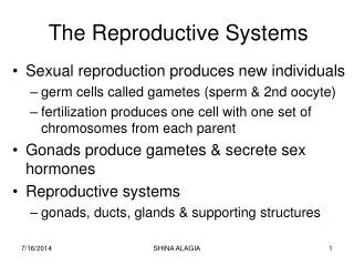
General Terminology: Gonads, or Primary Sex Organs = ovaries and testes Produce gametes & hormones Transportation System Transport of gametes Secondary or Accessory Organs Glands External genitalia
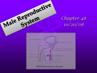
Male Reproductive Anatomy • Primary reproductive organs produce gametes • Secondary reproductive organs . . . • Male reproductive and urinary tracts are partially shared
Testes(paired gonads) 4 month • Develop adjacent to kidneys • Descend into scrotum through inguinal canal (function of gubernaculum testis) • Blood Supply via gonadal arteries • Peritoneal lining is carried along lining of scrotum • Spermatic cord: bundle containing all the “duct work”
Cryptorchidism In 3% of full-term and 30% of premature deliveries Significance? - Treatment?
External Features Function: supports, protects, and regulates temperature • Scrotum consists of Skin Dartos muscle Tunica vaginalis Median raphe • Involuntary contraction (cremasteric reflex) in response to cold or arousal
Internal Structure of Testes • Fibrous capsule – tunica albuginea – surrounds testes • Lobules contain approx. 800 Seminiferous Tubules collect at rete testis • Interstitial (Leydig) Cells make testosterone • Sustentacular (Sertoli) cells aid spermatogenesis
Spermatogenesis • Spermatogonia divide and one of the daughter cells matures into a primary spermatocyte. • Meiosis Spermatids (haploid). begins, 2 secondary spermatocytes. Another division • Spermiogenesis: Spermatid maturation into spermatozoa within sustentacular (Sertoli) cells • Spermiation: Spermatozoon released into lumen
Sustentacular (Sertoli) CellsInterstitial (Leydig) Cells Sertoli Cells: Maintenance of blood testis barrier special lumen fluid high in sex hormones, K+ and aa protection from immune attack (due tosperm specific Ag) Suspend spermatids and support spermatogenesis and spermiogenesis FSH and Testosterone work via Sertoli cellsSecretion of inhibin to slow sperm production Secretion of androgen-binding protein (ABP) Interstitial CellsLocated between tubulesproduce testosterone
Anatomy of a Spermatozoon Mature sperm has 3 portions: • Head with acrosome (containing enzymes) and compressed nucleus • Middle piece with lots of mitochondria. Why? • Tail - flagellum - (rotating in corkscrew fashion) See fig 27-6
Epididymis ~ 7 m long Head - superior, receives spermatozoa Body - distal and inferior Tail - leads to ductus deferens Functions: 1) Monitors and adjusts tubular fluid (lining has stereocilia!) 2) Recycles damaged spermatozoa 3) Stores sperm and facilitates maturation (capacitation) Rete testis and Efferent ductules
Spermatic Cord Can be palpated as it passes over the pubic brim. Constituents : 1. Pampiniform plexus of spermatic vein 2. Spermatic artery • Ductus (vas) deferens 4. Lymphatics 5. Nerves-ilioinguinal and genitofemoral Fig 27.3
Accessory Glands Provide for 95% of the seminal fluid 1) Seminal Vesicles • Paired, on back wall of urinary bladder • Tubular (~ 15 cm) • Produce 60% of semen, hormones, fructose, etc. • Activate sperm (leading to motility)
Prostate Gland 20 - 30% of seminal fluid Single, doughnut-shaped Secretion contains: • Citrate • Seminal plasmin (mild antibiotic) • Prostate specific antigen (PSA) – blood test for ?
Prostate Cancer • Usually grows slowly • Often slow urination is first sign • Digital rectal Exam and/or PSA • Treatment depends on size of tumor and other factors
Bulbourethral glands (Cowper’s glands) Pea size, paired, at base of penis Produce about 10% of semen Alkaline mucus buffers the acid that may be present in urine
2-5 ml ejaculate Ejaculation of semen by pelvic floor and penile muscles (Sympathetic division induces peristalsis in tract) Constituents: 1. sperm - 20 - 100 million sperm/ ml 2. seminal fluid – 60% from seminal vesicles 3. enzymes - proteases and seminalplasmin Semen
Penis Root - fixed to ischial rami Glans – enlargement of corpus spongiosum Prepuce = foreskin – partially covers glans and surrounds external urethral meatus (removed in circumcision) Preputial glands - produce smegma (supports bacterial growth, such as E. coli) Fig 27.9
Penis Shaft with erectile tissues Corpus Cavernosum Corpus Spongiosum Dorsal Artery and Vein Penile Urethra
Pathway of Sperm • Seminiferous tubules • Convoluted, then Straight • Rete testis • Efferent Ductules • Epididymis (head, body, tail) • Vas (ductus) deferens • Ampulla of vas deferens • Ejaculatory duct • Prostatic urethra • Membranous urethra • Penile (spongy) urethra Fig 27.8