The Reproductive Systems
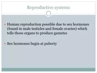
The Reproductive Systems. Sexual reproduction produces new individuals germ cells called gametes (sperm & 2nd oocyte) fertilization produces one cell with one set of chromosomes from each parent Gonads produce gametes & secrete sex hormones Reproductive systems

The Reproductive Systems
E N D
Presentation Transcript
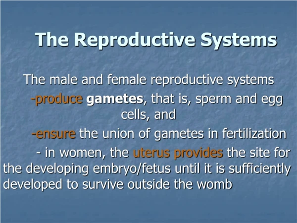
The Reproductive Systems • Sexual reproduction produces new individuals • germ cells called gametes (sperm & 2nd oocyte) • fertilization produces one cell with one set of chromosomes from each parent • Gonads produce gametes & secrete sex hormones • Reproductive systems • gonads, ducts, glands & supporting structures SHINA ALAGIA
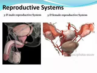
Male Reproductive System • Gonads, ducts, sex glands & supporting structures • Semen contains sperm plus glandular secretions SHINA ALAGIA
Scrotum • Sac of loose skin, fascia & smooth muscle divided into two pouches by septum • Skin contains dartos muscle causes wrinkling • Temperature regulation of testes • sperm survival requires 3 degrees lower temperature than core body temperature • cremaster muscle in spermatic cord • elevates testes on exposure to cold & during arousal • warmth reverses the process SHINA ALAGIA
Scrotal sac layers SHINA ALAGIA
Testes • Paired oval glands • Surrounded by dense white capsule called tunica albuginea • septa form 200 - 300 compartments called lobules • Each is filled with 2 or 3 seminiferous tubules where sperm are formed SHINA ALAGIA
Descent of Testes • Develop near kidney on posterior abdominal wall • Descends into scrotum by passing through inguinal canal • during 7th-8th month of fetal development SHINA ALAGIA
Tunica Vaginalis Tunica vaginalis • Piece of peritoneum that descended with testes into scrotal sac. • Allows for easier movement of testes within scrotum SHINA ALAGIA
Cryptorchidism • Testes do not descend into the scrotum • 3% of full-term & 30% of premature infants • Untreated bilateral cryptorchidism results in sterility & a greater risk of testicular cancer • Descend spontaneously 80% of time during the first year of life • surgical treatment necessary before 18 months SHINA ALAGIA
Stages of Sperm Formation • Seminiferous tubules contain • all stages of sperm development: spermatogonia, primary spermatocyte, secondary spermatocyte, spermatid, spermatozoa • supporting cells called sertoli cells • Leydig cells in between tubules secrete testosterone SHINA ALAGIA
Supporting Cells • Sertoli cells -- extend from basement membrane to lumen • form blood-testis barrier • Secrete ABP (Androgen binding protein) • secrete inhibin which slows sperm production by inhibiting FSH SHINA ALAGIA
Epididymis • Comma-shaped organ, 1.5in long along posterior border of each testis • Head, body and tail region • Multiple efferent ducts become a single ductus epididymis in the head region • 20 foot tube if uncoiled • Tail region continues as vas deferens SHINA ALAGIA
Histology of the Epididymis • Ductus epididymis • lined with pseudostratified ciliated columnar epithelium (stereocilia) • Site of sperm maturation • motility increases over 2 week period • Storage for 1-2 months • Propels sperm onward SHINA ALAGIA
Ductus (Vas) Deferens • Pathway of 18 inch muscular tube • ascends along posterior border of epididymis • passes up through spermatic cord and inguinal ligament • reaches posterior surface of urinary bladder • empties into prostatic urethra with seminal vesicle • Lined with pseudostratified columnar epithelium & covered with heavy coating of muscle • convey sperm along through peristaltic contractions • stored sperm remain viable for several months SHINA ALAGIA
Spermatic Cord • All structures passing to and from the testes • testicular artery • pampiniform plexus of veins • autonomic nerves • lymphatic vessels • ductus (vas) deferens • cremaster muscle SHINA ALAGIA
Inguinal Canal & Inguinal Hernias • Inguinal canal is 2 inch long tunnel passing through the 3 muscles of the anterior abdominal wall -- weakens wall • originates at deep inguinal ring and ends at superficial ring • Indirect hernia -- loop of intestine protruding through deep ring • Direct hernia -- loop of intestine pushes through posterior wall of inguinal canal • More common in males SHINA ALAGIA
Inguinal Canal SHINA ALAGIA
Ejaculatory Ducts • Formed from duct of seminal vesicle & ampulla of vas deferens • Adds fluid to prostatic urethra just before ejaculation SHINA ALAGIA
Urethra • 8 inch long passageway for urine & semen • Prostatic urethra (1 inch long) • Membranous urethra (passes through UG diaphragm ) • Penile (spongy) urethra (through corpus spongiosum) SHINA ALAGIA
Seminal Vesicles • Pair of pouchlike organs found posterior to the base of bladder • Alkaline, viscous fluid • neutralizes vaginal acid & male urethra • fructose for ATP production • clotting proteins for coagulation of semen Posterior View SHINA ALAGIA
Prostate Gland • Single organ the size of chestnut found inferior to bladder • Secretes milky, pH 6.5 fluid that increases sperm motility and viability • citric acid for ATP production & enzymes for seminal liquefaction SHINA ALAGIA
Bulbourethral or Cowper’s Gland • Paired, pea-sized gland within the UG diaphragm • Secretes alkaline mucous into spongy urethra • Neutralizes acids and lubricates SHINA ALAGIA
Penis • Passageway for semen & urine • Body composed of three erectile tissue masses filled with blood sinuses • Composed of bulb, crura, body & glans penis SHINA ALAGIA
Cross-Section of Penis • Corpora cavernosa • upper paired, erectile tissue masses • Corpus spongiosum • lower erectile tissue mass • surrounds urethra • ends as glans penis SHINA ALAGIA
Root of Penis & Muscles of Ejaculation • Bulb of penis or base of corpus spongiosum enclosed by bulbospongiosus muscle • Crura of penis or ends of corpora cavernosa enclosed by ischiocavernosus muscle SHINA ALAGIA
Glans Penis • Enlarged distal end of corpus spongiosum • External urethral orifice is small slit • Covered by loosely fitting prepuce or foreskin SHINA ALAGIA
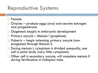
Female Reproductive System • Ovaries produce 2nd oocytes & hormones • Uterine tubes transport fertilized ova • Uterus where fetal development occurs • Vagina & external genitalia constitute the vulva • Mammary glands produce milk SHINA ALAGIA
The Ovary • Pair of organs, size of unshelled almonds found in upper pelvic region • Regional histology • tunica albuginea is capsuleof dense connective tissue • cortex is region just deep totunica, contains follicles • medulla is deeper regioncomposed of connective tissue, blood vessels & lymphatics • germinal epithelium is simple epithelial covering over the ovary SHINA ALAGIA
Ovary SHINA ALAGIA
Reproductive Ligaments • Broad ligament suspends uterus from side wall of pelvis • Mesovarium attaches ovaries to broad ligament • Ovarian ligament anchors ovary to uterus • Suspensory ligament covers blood vessels to ovaries • Round ligament attaches ovaries to inguinal canal SHINA ALAGIA
Follicular Stages • Stages of follicular development • primordial • primary • secondary • graafian • ovulation • Corpus luteum is ovulation wound • fills in with hormone secreting cells • Corpus albicans is white scar left after corpus luteum is not needed SHINA ALAGIA
Histology of a Graafian Follicle • Zona pellucida -- clear area between oocyte & granulosa cells • Corona radiata is granulosa cells attached to zona pellucida--still attached to oocyte at ovulation • Antrum formed by granulosa cells secreting fluid • By this time, the oocyte has reached the metaphase of meiosis II stage and stopped developing -- first polar body has been discarded SHINA ALAGIA
Uterine or Fallopian Tubes • Narrow, 4 inch tube extends from ovary to uterus • infundibulum is open, funnel-shaped portion near the ovary • fimbriae are moving finger-like processes • ampulla is central region of tube • isthmus is narrowest portion joins uterus SHINA ALAGIA
Function of Uterine Tube • events occurring in the uterine tube • fimbriae sweep oocyte into tube, cilia & peristalsis move it along, sperm reaches oocyte in ampulla, fertilization occurs within 24 hours after ovulation & zygote reaches uterus about 7 days after ovulation SHINA ALAGIA
Anatomy of the Uterus • Site of menstruation& development of fetus • Description • 3 inches long by 2 in. wide and 1 in. thick • subdivided into fundus,body, isthmus & cervix • interiorly contains uterine cavity accessed by cervical canal (internal & external os) SHINA ALAGIA
Position of Uterus • Anteflexion -- normally projects anteriorly and superiorly over the urinary bladder • Retroflexion -- posterior tilting of the uterus SHINA ALAGIA
Histology of the Uterus • Endometrium • stroma of connective tissue and endometrial glands • stratum functionalis shed during menstruation • stratum basalis replaces stratum functionalis each month • Myometrium • 3 layers of smooth muscle • Perimetrium • visceral peritoneum SHINA ALAGIA
Vagina • Passageway for birth, menstrual flow & intercourse • 4 inch long fibromuscular organ ending at cervix • mucosal layer • stratified squamous epithelium & areolar connective tissue • large stores of glycogen breakdown to produce acidic pH • muscularis layer is smooth muscle allows considerable stretch • adventitia is loose connective tissue that binds it to other organs • lies between urinary bladder and rectum • orifice partially closed with membrane (hymen) SHINA ALAGIA
Vulva (pudendum) • Mons pubis -- fatty pad over the pubic symphysis • Labia majora & minora -- folds of skin encircling vestibule where find urethral and vaginal openings • Clitoris -- small mass of erectile tissue • Bulb of vestibule -- masses of erectile tissue just deep to the labia on either side of the vaginal orifice SHINA ALAGIA
Perineum • Diamond-shaped area between the thighs in both sexes • bounded by pubic symphysis and coccyx • urogenital triangle contains external genitals • anal triangle contains anus SHINA ALAGIA
Mammary Glands • Modified sweat glands • amount of adipose determines size of breast • milk-secreting glands open by lactiferous ducts at the nipple • areola is pigmented area around nipple • suspensory ligaments suspend breast SHINA ALAGIA
Fibrocystic Disease of the Breasts • Most common cause of breast lumps • Cysts and thickenings of alveoli develop • Cause • hormonal imbalance • excess of estrogen or deficiency of progesterone in the postovulatory phase • result is lumpy, swollen & tender breast a week before menstruation begins SHINA ALAGIA
Reproductive Physiology • Somatic cells (diploid cells) • 23 pairs of chromosomes for a total of 46 • each pair is homologous • one member of each pair is from each parent • 22 autosomes & 1 pair of sex chromosomes • sex chromosomes are either X or Y • females have two X chromosomes • males have an X and a smaller Y chromosome • Gametes (haploid cells) • single set of chromosomes for a total of 23 • produced by special type of division: meiosis SHINA ALAGIA
tetrad Meiosis I -- Prophase I • Chromosomes become visible, mitotic spindle appears, nuclear membrane & nucleoli disappear • Events not seen in prophase of Mitosis or Meiosis II • synapsis • all copies of homologous chromosomes pair off forming a tetrad • crossing-over • portions of chromatids are exchanged between any members of the tetrad and genetic recombination produces gametes unlike either parent SHINA ALAGIA
Exchange of Genetic Material • Chromosomes are exchanged between chromatids on homologous chromosomes SHINA ALAGIA
Spermatogenesis • Spermatogonium (stem cells) give rise to 2 daughter cells by mitosis • One daughter cell kept in reserve -- other becomes primary spermatocyte • Primary spermatocyte goes through meiosis I • DNA replication • tetrad formation • crossing over SHINA ALAGIA
Spermatogenesis- 2 • Secondary spermatocytes are formed from primary spermtocytes • 23 chromosomes of which each is 2 chromatids joined by centromere • goes thru’ meiosis II • 4 spermatids are formed • each is haploid & unique • all 4 remain in contact with cytoplasmic bridge SHINA ALAGIA
Spermiogenesis • Spermiogenesis = maturation of spermatids into spermatozoa (head body and tail differentiation) SHINA ALAGIA
Sperm Morphology • Adapted for reaching & penetrating a secondary oocyte • Head contains DNA & acrosome (hyaluronidase and protease enzymes) • Midpiece contains mitochondria to form ATP • Tail is flagellum used for locomotion SHINA ALAGIA
Hormonal Control of Spermatogenesis • Puberty • hypothalamus increases its stimulation of anterior pituitary with releasing GnRH • anterior pituitary increases secretion LH & FSH • LH stimulates Leydig cells to secrete testosterone • FSH stimulates spermatogenesis • with testosterone, stimulates sertoli cells to secrete androgen-binding protein (keeps hormones levels high) • testosterone stimulates final steps of spermatogenesis SHINA ALAGIA
Effects of Testosterone • Testosterone & DHT bind to receptors in cell nucleus & change genetic activity • Prenatal effect is born a male • At puberty, final development of 2nd sexual characteristics and adult reproductive system • sexual behavior & libido • male metabolism (bone & muscle mass heavier) • deepening of the voice SHINA ALAGIA