Download
1 / 59
590 likes | 610 Vues
Airway Management, Ventilation, Oxygen Therapy. Respiratory Anatomy. Nose and mouth (warms, moistens, and filters air). Pharynx Oropharynx Nasopharynx Epiglottis Trachea (windpipe). Respiratory Anatomy. Cricoid cartilage (adams apple). Larynx (voice box). Bronchi Lungs

E N D
Airway Management, Ventilation, Oxygen Therapy www.rxdentistry.net
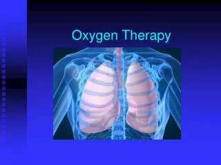
Respiratory Anatomy • Nose and mouth (warms, moistens, and filters air). • Pharynx • Oropharynx • Nasopharynx • Epiglottis • Trachea (windpipe) www.rxdentistry.net
Respiratory Anatomy • Cricoid cartilage (adams apple). • Larynx (voice box). • Bronchi • Lungs • Visceral pleura (surface of lungs) • Parietal pleura (internal chest wall) • Interpleural space (potential space) www.rxdentistry.net
Respiratory Anatomy • Diaphragm • Inhalation (active process) • Diaphragm and intercostal muscles contract, increasing the size of the thoracic cavity. • Diaphragm moves slightly downward, ribs move upward and outward. • Air flows into the lungs creating a negative pressure in the chest cavity. www.rxdentistry.net
Respiratory Anatomy • Exhalation (passive process) • Diaphragm and intercostal muscles relax decreasing the size of the thoracic cavity. • Diaphragm moves upward, ribs move downward and inward. • Air flows out of the lungs creating a positive pressure inside the chest cavity. www.rxdentistry.net
Respiratory Physiology • Oxygenation - blood and the cells become saturated with oxygen • Hypoxia - inadequate oxygen being delivered to the cells • Signs of Hypoxia • Increased or decreased heart rate • Altered mental status (early sign) • Agitation • Initial elevation of B.P. followed by a decrease • Cyanosis (often a late sign) www.rxdentistry.net
Alveolar/Capillary Exchange • Oxygen-rich air enters the alveoli during each inspiration. • Oxygen-poor blood in the capillaries passes into the alveoli. • Oxygen enters the capillaries as carbon dioxide enters the alveoli. www.rxdentistry.net
Capillary/Cellular Exchange • Cells give up carbon dioxide to the capillaries. • Capillaries give up oxygen to the cells. www.rxdentistry.net
Infant and Child Considerations • Mouth and nose - generally all structures are smaller and more easily obstructed than in adults. • Pharynx - infant’s and children’s tongues take up proportionally more space in the mouth than adults. • Trachea - (windpipe) • Infants and children have narrower tracheas that are obstructed more easily by swelling. • Trachea is softer and more flexible in infants and children. www.rxdentistry.net
Infant and Child Considerations • Cricoid cartilage - like other cartilage in the infant and child, the cricoid cartilage is less developed and less rigid. It is the narrowest part of the infant’s or child’s airway. • Diaphragm - chest wall is softer, infants and children tend to depend more heavily on the diaphragm for breathing. www.rxdentistry.net
Opening the Mouth • Crossed-finger technique • Inspect the mouth • Vomit • Blood • Secretions • Foreign bodies • Be extremely cautious • Fingers • Gag or vomit www.rxdentistry.net
Opening the Airway • Head-tilt, chin lift maneuver • Adults vs.. Infants and Children • Jaw thrust maneuver www.rxdentistry.net
Techniques of Suctioning • BSI precautions • Purpose • Remove blood, other liquids, and food particles from the airway • Some suction units are inadequate for removing solid objects like teeth, foreign bodies, and food • A patient needs to be suctioned immediately when a gurgling sound is heard with artificial ventilation www.rxdentistry.net
Types of Suction Units • Mounted Suction Devices • Fixed on-board the ambulance • 300mmHg pull on gauge when tubing is clamped • Should be adjustable for infants and children • Powered by ambulance engine manifold www.rxdentistry.net
Portable Suction Devices • Electric - battery powered • Oxygen - powered • Hand - powered • Each device must have • Wide-bore, thick walled, non-kink tubing • Plastic collection bottle, supply of water • Enough vacuum to clear the throat www.rxdentistry.net
Suction Catheters • Hard or rigid catheter (Yankaeur) • Tonsil tip • Used to suction mouth and oropharynx • Inserted only as far as you can see • Use extreme caution on infants and children • Soft tissue damage www.rxdentistry.net
Suction Catheters • Soft catheter (French catheter) • Nose or nasopharynx, mouth • Measured from tip of the nose to the tip of his ears. • Not inserted beyond the base of the tongue www.rxdentistry.net
Techniques of Suctioning • Positioned at patient’s head • Turn on the suction unit • Select catheter • Measure and insert without suction if possible • Suction from side to side • Adults no more than 15 seconds • Infants & children no more than 5 seconds • Rinse catheter with water if necessary www.rxdentistry.net
Special Considerations • Secretions that cannot be removed log roll and finger sweep • Patient producing frothy secretions as rapidly as suctioning can remove them • Suction 15 seconds • Positive pressure with supplemental oxygen for 2 minutes then suction again and repeat the process • Residual air removed from lungs, monitor pulse and heart rate • Before and after suctioning hyperventilate 24 per/min. x 5 min. www.rxdentistry.net
Oropharyngeal Airway (OPA) • Used to maintain a patent airway only on deeply unresponsive patients • No gag reflex • Designed to allow suctioning while in place • Must have the proper size • If patient becomes responsive and starts to fight the OPA remove it... www.rxdentistry.net
Inserting the OPA • Select the proper size (corner of the mouth to tip of the ear) • Open the patient’s mouth • Insert the OPA with the tip facing the roof of the mouth • Advance while rotating 180° • Continue until flange rests on the teeth • Infants and children insertion www.rxdentistry.net
Nasopharyngeal Airway (NPA) • Nose hose, nasal trumpet • Used on patients who are unable to tolerate an OPA or is not fully responsive • Do not use on suspected basilar skull fracture • Still need to maintain head-tilt chin lift or jaw thrust when inserted • Must select the proper size • Made to go into right nare or nostril www.rxdentistry.net
Inserting the NPA • Select the proper size in length and diameter • Lubricate • Insert into right nostril with bevel always toward the septum • Continue inserting until flange rests against the nostril • Insertion into left nostril www.rxdentistry.net
Assessment of Breathing • After establishing an airway your next step should be to assess breathing • Look • Breathing pattern regular or irregular • Nasal flaring • Adequate expansion, retractions www.rxdentistry.net
Assessment of Breathing • Listen • Dyspnea when speaking • Unresponsive place ear next to patients mouth • Is there any movement of air? www.rxdentistry.net
Assessment of Breathing • Feel • Check the volume of breathing by placing you ear and cheek next to the patient’s mouth www.rxdentistry.net
Assessment of Breathing • Auscultate • Stethoscope • Mid clavicular about the second intercostal space and the fourth or fifth anterior midaxillary line or next to sternum • Check both sides • Present and equal bilaterally • Diminished or absent www.rxdentistry.net
Adequate Breathing • Normal rate • Adult 12 - 20/min • Child 15 - 30/min • Infant 25 - 50/min • Rhythm • Regular • Irregular www.rxdentistry.net
Adequate Breathing • Quality • Breath sounds present and equal • Chest expansion adequate and equal • Effort of breathing • use of accessory muscles predominately in infants and children • Depth • Adequate chest rise and fall • Full breath sounds heard www.rxdentistry.net
Inadequate Breathing • Rate • Outside the normal limits • Tachypnea (rapid breathing) • Badypnea (slow breathing) • Rhythm • Irregular breathing pattern www.rxdentistry.net
Inadequate Breathing • Quality • Breath sounds diminished or absent • Excessive use of accessory muscles, retractions • Diaphormatic breathing • Nostril flaring (infants & children) • Depth • Shallow breathing • Agonal respirations - occasional gasping respirations • Any of these signs is by itself is a reason to ventilate a patient without delay www.rxdentistry.net
Positive Pressure ventilation • The practice of artificially ventilating, or forcing air into a patient who is breathing inadequately or not breathing at all www.rxdentistry.net
Techniques of Artificial Ventilation • In order of preference • Mouth to mask • Two-person bag-valve-mask • Flow-restricted oxygen-powered ventilation device • One-person bag-valve-mask www.rxdentistry.net
Considerations When Using Artificial Ventilation • Maintain a good mask seal • Device must deliver adequate volume of air to sufficiently inflate the lungs • Supplemental oxygen must be used www.rxdentistry.net
Adequate Artificial Ventilations • Chest rises and falls with each ventilation • Rate of ventilations are sufficient • Heart rate returns to normal • Color improves www.rxdentistry.net
Inadequate Artificial Ventilations • Chest does not rise and fall • Ventilation rate is too fast or slow • Heart rate does not return to normal www.rxdentistry.net
Mouth-to-Mouth Ventilation • Air we breath contains 21% oxygen • 5% used by the body • 16% is exhaled • Danger of infectious disease www.rxdentistry.net
Mouth-to-Mask • Eliminates direct contact with patient • One-way valve system • Can provide adequate or greater volume than a BVM • Oxygen port (should be connected to 15 lpm) www.rxdentistry.net
Bag-Valve-Mask (BVM) • EMT-B can feel the lung compliance • Consists of self-inflating bag, one-way valve, face mask, intake/oxygen reservoir valve, and an oxygen reservoir. • By adding oxygen and a reservoir close to 100% oxygen can be delivered to the patient • When using a BVM an OPA/NPA should be used if possible www.rxdentistry.net
Bag-Valve-Mask Cont... • Volume of approximately 1,600 milliliters • Provides less volume than mouth-to-mask • Single EMT may have trouble maintaining seal • Two EMT’s more effective • Pop-off valve must be disabled • Available in infant, child, and adult sizes www.rxdentistry.net
Flow-Restricted, Oxygen-Powered Ventilation Device • Known as a demand-valve device • Can be operated by patient or EMT • Unable to feel lung compliance • With proper seal will deliver 100% oxygen • Designed for use on adult patients • Gastric distension • Rupture of the lungs • A trigger positioned to allow EMT to keep both hands on the mask www.rxdentistry.net
Automatic Transport Ventilators • Deliver 100% oxygen • Provide and maintain a constant rate and tidal volume during ventilation • Advantages • Frees both hands • Rate, & tidal volume can be set • Alarm for low oxygen tank • Disadvantages • Oxygen powered • not used in children under 5 • Cannot feel increase in airway resistance www.rxdentistry.net
Oxygen Therapy • Oxygen is a drug that can be given by the EMT-B • “Generally speaking”, a patient who is breathing less than 12 and more than 24 times a minute needs oxygen of some kind www.rxdentistry.net
Oxygen Dangers • Oxygen supports combustion, (it is not flammable) • Avoid contact with petroleum products • Smoking • Handle carefully since contents are under pressure • Strap the cylinder between the patients legs on the cot so it doesn’t fall www.rxdentistry.net
Oxygen Cylinders • All of the cylinders when full are the same pressure of 2,000 psi. • Usually green or aluminum grey • D cylinder - 350 liters • E cylinders - 625 liters • M cylinders - 3,000 liters • G cylinders - 5,300 liters • H cylinders - 6,900 liters www.rxdentistry.net
High-Pressure Regulator • Provides 50 psi to an oxygen-powered, ventilation device. • Flow rate cannot be controlled www.rxdentistry.net
Low Pressure/Therapy Regulator • Permit oxygen delivery to the patient at a desired rate in liters per minute • Flow rate can go from 1 to 25 liters/min. www.rxdentistry.net
Oxygen Humidifiers • Dry oxygen is not harmful in the short term • Generally not needed in prehospital care • Transport time of an hour or more humidifier should be considered www.rxdentistry.net
Changing Oxygen Bottle • Check cylinder for oxygen remove protective seal • Quickly open and shut tank to remove debris • Place regulator over yoke and and align pins. • Make sure new O ring is in place • Hand tighten the T screw • Open to check for leaks www.rxdentistry.net
Nonrebreather Mask • Preferred method of giving oxygen to prehospital patients • Up to 90% oxygen can be delivered • Bag should be filled before placing on patient • Flow rate should be adjusted to 15 liters/min. • Patients who are cyanotic, cool, clammy or short of breath need oxygen • Concerns of too much oxygen • Different size masks www.rxdentistry.net