Impact of TGF-β1 on cPML Expression in HepG2 and MEF Cell Lines
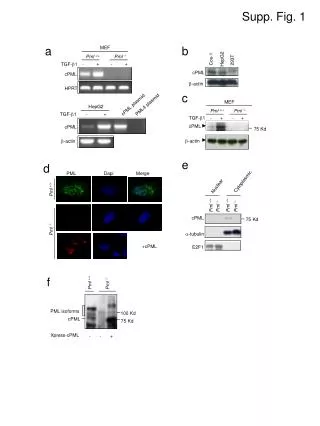
This study investigates the roles of cPML isoforms in HepG2 and MEF cell lines under the influence of TGF-β1. Using various plasmids and cell lines, we examine the expression levels of cPML, β-actin, and E2F1. We analyze the localization of cPML in both cytoplasmic and nuclear compartments, observing different behavior in Pml.+/+ and Pml.-/- conditions. The results indicate a complex relationship between cPML expression and TGF-β1 stimulation, shedding light on the regulatory mechanisms involved in tumorigenesis and cellular response.

Impact of TGF-β1 on cPML Expression in HepG2 and MEF Cell Lines
E N D
Presentation Transcript
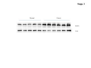
Supp. Fig. 1 a b MEF Pml +/+ Pml -/- HepG2 Cos-1 293T + + TGF-1 - - cPML cPML -actin HPRT c MEF cPML plasmid PML4 plasmid HepG2 Pml +/+ Pml -/- TGF-1 - + TGF-1 - + - + cPML cPML 75 Kd -actin -actin e d PML Dapi Merge Cytoplasmic Nuclear Pml +/+ Pml +/+ Pml -/- Pml -/- Pml +/+ cPML 75 Kd Pml -/- -tubulin E2F1 +cPML f Pml +/+ Pml -/- PML isoforms 100 Kd cPML 75 Kd Xpress-cPML - - +