Download
1 / 42
430 likes | 651 Vues
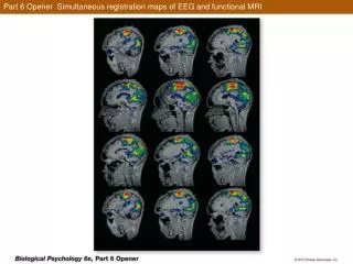
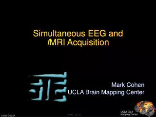
Recording of EEG by MRI. Your scanner can measure more than you think. Lars G. Hanson MR-physicist Danish Research Centre for MR DRCMR, Copenhagen. Electrophysiology: ECG, EEG, EMG,... Measurement of electrical activity in the body. High temporal, low spatial resolution.

E N D
Recording of EEG by MRI Your scanner can measure more than you think Lars G. Hanson MR-physicist Danish Research Centre for MR DRCMR, Copenhagen
Electrophysiology: ECG, EEG, EMG,... • Measurement of electrical activity in the body. • High temporal, low spatial resolution. • EEG measured during fMRI: • fMRI can supplement EEG, e.g., for localisation of epileptic foci: Timing from EEG, position from BOLD. • However: Like using phone in a thunder storm. • Periods of silence followed by extreme electrical noise and distortion. • ”Thunder bolts” arrive every millisecond!
Overview of talk • Incentive for doing EEG-fMRI and complications regarding interpretation. • Technical challenges: • Field-pulse-interactions (ballistocardiac EEG distortions) • EEG distortions induced by imaging • A solution: Using the scanner for EEG-recording
fMRI • fMRI for functional mapping: • Intensity variations over time reflect activity due to the BOLD effect: • The oxygen consumption increases. • The oxygen supply increases more. • Deoxy-haemoglobin is paramagnetic (decreases the MR signal). • Hence BOLD is a very indirect measure of activity, and the temporal resolution is bad. • fMRI can only tell where activity is, if we know when it occurs (unrelated changes dominate).
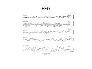
EEG • EEG is a more direct measure of neuronal activity, and the temporal resolution is excellent. • Certain brain states can only be detected by EEG, e.g. sleep staging, epileptic spikes. • The spatial resolution, however, is quite limited. If EEG can answer the ”when”-question, can fMRI then answer the ”where”-question?
Why EEG-fMRI? Example: Localizing foci in epilepsy. • Inter-ictal spiking activity can be registered by EEG. Correlation to fMRI time series can reveal where corresponding BOLD response is present. Hypothesis: Correlation maps give the foci locations needed for presurgical planning. However…
Epilepsy EEG-fMRI complications • BOLD response may appear at the source of the spiking activity. • BOLD response may appear elsewhere as a result of the spiking activity. • The haemodynamic response may be different in epileptogenic zones. Oxygen consumption may, for example, be balanced by supply, i.e., NO BOLD.
EEG-fMRI potential Despite interpretation challenges, EEG-fMRI is a very promising technique: • Increased spatial resolution of EEG and temporal resolution of fMRI. • EEG-fMRI may reveal causality. • EEG be used to monitor alertness/brain state in fMRI-experiments, e.g., sleep staging. • EEG provides classification of single events, e.g., response time or level of surprise. Effects normally lost in fMRI averaging may be observable (e.g. Debener et al, ’06)
Overview of talk • Incentive for doing EEG-fMRI and complications. • Technical challenges: • Field-pulse-interactions (ballistocardiac EEG distortions) • Imaging induced EEG distortions • A solution: Using the scanner for EEG-recording
Field-pulse interactions Effect of a high magnetic field on EEG: • Pulse contributions are amplified and changed. • The “ballisto-cardiac effect”: Voltages are induced orthogonally to blood stream. • Minor electrode movement give significant noise. These effects are present even without imaging. Imaging adds overwhelming extra problems…
Spatially and temporally varying magnetic field. Changes every milisecond
Spatially and temporally varying magnetic field. Changes every milisecond
Typical solution • Imaging gradient induced noise is repeated. • Can be averaged over a number of images and be subtracted from the subsequent images (template subtraction, Allen et al, 2001). • Large signal – Large artefact = tiny EEG. Problematic: • Requires microsec. timing, excellent linearity and pt. must be motionless. • Average too long, and the patient moves. Too short, and the EEGs are erroneous. • Much better to avoid gradient artifacts in the first place!
Overview of talk • Incentive for doing EEG-fMRI and complications. • Technical challenges: • Field-pulse-interactions (ballistocardiac EEG distortions) • Imaging induced EEG distortions • Avoiding imaging artifacts by using the scanner for EEG-recording
EPI sequence: Anami: Decent EEG can be measured in silent periods. MR signal is recorded in the same periods!
Idea: “Magstripe” MRI The physiological signals are modulated and emitted as radio waves that are measured by the scanner along with the MR-signal. ”Soundtrack” saved next to image.
Spectrogram of recorded signal Milli-second time resolution
Motivation • Imaging induced EEG-artifacts are largely avoided. • Microsecond synchronisation is inherent. • Needed for removal of residual artefacts from e.g. eddy currents (Cohen, 01) • Inexpensive and easy: • Surplus bandwidth of the scanner is employed for non-MR purposes. • No need for MR compatible EEG acquisition system. Signal paths reused. • MRI- and EEG-data meet in acquisition computer. • Facilitates PACS storage and EEG/fMRI correlation. • Robust: • Traditional filtering relies on subtraction of large signals to isolate EEG.
Pilot study, day 1 Amplitude modulated RF RF transmitter Signal “FID” recorded by scanner
Fully featured 8-ch EEG system for MRI Gradient sensor Antenna Electrodes Frequency-converter and amplitude-modulator Amplifier
MR movie and extracted electrophysiology ECG EOG Calibration signal Movie is based on old hardware: Illustrative, but noise is excessive
New implementation • 8 channel amplitude modulator. • 0-130 MHz in 0,1 Hz steps (digital synthesis) • Frequencies, filters and timings are programmable via PC. • Online viewing of recorded signals at all times. • Design and implementation by Brother Christian.
MRI Oversampling • Stripes in images interfere with postprocessing. • Solution provided by scanner vendors: Modern scanners measure images wider than prescribed. Signal at oversampled frequencies is discarded during image reconstruction. Hence the method does not interfere with any imaging method. Capacity for at least 60 channels.
Movie acquired with new hardware 2xEOG and 2xEEG (F3-F4) Oversampling is here used to avoid imaging artefacts
EEG EOG EOG EEG slc1 slc2 slc3 Correlation maps, full bandwidth
Encoding of electrophysiology in fMRI timeseries F3-F4 Temporal corr. fMRI timeseries
EEG from F3-F4 Only pulse and eye motion artefacts are clearly recognisable features in this recording Pulse artefacts
Typical concerns • Encoded signals will interfere with postprocessing. • This will disturb non-linear image reconstruction. • This technique will not work with spiral EPI and other ramp-sampling sequences. • Extra noise will be added to EEG. • Non-MR takes dynamic range from MR signals. • Scanner bandwidth is needed for EPI. • Sampling is non-equidistant. Valid concerns - but dry your eyes. Go see http://www.MRIware.com/
0 1 2 3 4 5 Alpha recording • Recordings after removal of residual gradient artifacts: Note: Pulse artifacts dominate all channels.
Alpha recording • Spectrogram after pulse artifact removal using fMRIB-plugin for EEGlab (Nieazy et al, 2005): Alpha Paradigm: Eyes closed-open-closed, 30 secs. each
The prototype • 8 channel modular design • Programmable via serial interface (carrier frequencies, trigger timings, filter bandwidths,…) • Real-time previewing via serial interface (low quality compared to recording by scanner).
Summary, EEG-fMRI • Epilepsy was driving the EEG-fMRI field, but clinical application is problematic. • However, EEG-fMRI significantly aids interpretation and adds specificity to fMRI. It also provides entirely new possibilities. • The technical challenges, pulse and imaging induced EEG-distortions can be handled, e.g., by recording EEG with scanner.
Summary, Magstripe MRI • Your scanner is a very capable piece of hardware. You paid a lot for it. You might as well use it for everything. • Gradient artifacts are largely avoided and residuals easily filtered. Adds robustness. • The method and prototype is easy to use and works well. Comparison is pending. • Generic technique: Any signal, any scanner, but raw data access is needed.
Avoidance of artifacts Precise frequency is needed. Must match bpp:
Typical concerns • This will disturb non-linear image reconstruction. • Stripes in images will interfere with my post-processing. Signal at oversampled frequencies is discarded early (OS is default). Normally pure noise that should not affect reconstruction.
Typical concerns Concern: Scanner bandwidth is needed for EPI. • Siemens Trio has 8 MHz bandwidth (8 channels x 1 MHz) • Typically 200 kHz is used for EPI. • 60 EEG channels with 10 kHz bandwidth are easily fit in, • also when doing parallel imaging. • Additional 700 with wiring to coil interface. • Only 420 if parallel imaging is performed. • Why all this excess bandwidth? • Filter edge-effects occur but are not much of a concern.
Typical concerns Concern: This technique depends on periods free of gradient acticity and will not work with e.g. spiral EPI and other ramp-sampling sequences. • It does: • Perfect syncronization facilitates gradient artifact removal. • 3 stages in gradient artifact removal (artifact is 10 times signal):
Typical concerns • Extra noise will be added to my EEG. • Non-MR takes dynamic range from MR signals • Sure, but these effects are insignicant: • MR signal is very localized in time. • Non-MR is evenly distibuted over time. • MR: Well-localized in k-space. • Non-MR: Well-localized in image space. • Optimal situation: • - Even low-amplitude RF give high SNR.
New implementation • 8 channel amplitude modulator. • 0-130 MHz in 0,1 Hz steps (digital synthesis) • Frequencies, filters and timings are programmable via PC. • Online viewing of recorded signals at all times. • Design and implementation by Brother Christian.
The gradient trigger • Gradient artifacts have high bandwidth and amplitude. Requires more bandwidth than EEG and may saturate amplifier. Hence the gradient trigger.