Download

1 / 21
210 likes | 320 Vues
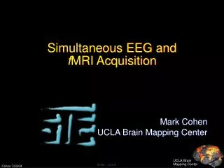
This study explores the use of simultaneous MRI and EEG to detect interictal epileptic activity, overcoming the limitations of each method individually. By tightly interleaving the acquisition of EEG and MRI data, researchers were able to identify neural activity patterns and correlate them with fMRI responses. Results from a case study of a 43-year-old man with right mesial temporal lobe epilepsy showed agreement between EEG data, fMRI maps, and pre-operative diagnosis, leading to successful post-operative outcomes. Acknowledging the challenges of variability in MR temporal courses and the need for further data averaging, this research highlights the promising potential of integrated MRI and EEG technology in epilepsy diagnosis and treatment.

E N D
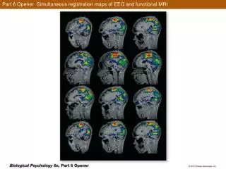
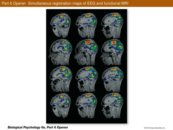
Evidence of Interictal Epilepctic Activity Using Simultaneous MRI and EEG
Alberto L. Vazquez, M.S.* Steve B. Baumann, Ph.D.+ Mark L. Scheuer, M.D.+ Douglas C. Noll, Ph.D.* * Dept. Biomedical Engineering University of Michigan, Ann Arbor, MI + University of Pittsburgh Medical Center, Pittsburgh, PA
Background • A disadvantage of fMRI responses is its sluggish temporal characteristics while a disadvantage of EEG is its poor spatial localization • Combination of EEG and MRI should provide a powerful tool, especially when stimulus cannot be induced, measured, and/or controlled
Background • Problems of integration • gradient-induced currents • saturation of pre-amplifier/electronics • movement and pulsatile flow induced currents • RF artifact due to shielding effects of conductors • susceptibility artifacts from electrodes • safety issues
Background • Variety of methods exist to acquire EEG and MRI data • trigger MR with EEG activity • Interleave EEG and MRI (different methods of interleaving) • Our goal: acquire EEG and MRI tightly interleaved (with minimal loss of EEG signals) throughout a scan
EEG • MRI compatible cap (Electrode Cap, Inc.) • Ear used as EEG voltage reference • 64 channels referentially recorded (uninterpretable) • Bipolar reconstruction (interpretable) • Data band-pass filtered 0.1-100 Hz
EEG • EEG acquired while RF and gradients are quiet • Data sampled at 256 Hz • Closely interleaved acquisition 100ms 100ms 100ms 650ms 650ms …… t MR EEG MR MR EEG slice 1 slice 3 slice 2
MRI • GE Signa 1.5 T scanner • Single-shot EPI (even slice spacing) TR = 3000ms TE = 40ms FA = 90deg • 4 axial slices (7mm, skip 3mm) centered about right temporal lobe
MRI • Resolution = 3.125mm x 3.125mm • Scan duration = 30mins • Reference used Gamma function • Delay = 2s • Data window of 1min (trends, …) 1 min
Subject • 43 year-old man • Right mesial temporal lobe epilepsy • Resting EEG contained right anterior temporal sharp waves • MR images show evidence of right mesial temporal sclerosis • (image???)
Results ROI r > 0.2, p < 0.15
Results MRIData Sharp Wave temporal electrode temporal electrode
Results Presumedhead movement temporal electrode posterior electrode posterior electrode
Results posterior electrode
Discussion • EEG data shows neural activity ~300ms in duration • fMRI response lasted ~12s (delay to onset = 2s) • fMRI map agrees with pre-operative diagnosis • Post-operative findings showed temporal mesial sclerosis in resected tissue (subject has been 8 months seizure-free)
Discussion • MR temporal course shows large amount of variability • Subject motion • Little averaging due to small number of detected events (n=2) • Physiological noise, ... • No subject motion artifacts were present in correlation map
Acknowledgements • Electrode Cap, Inc. (???) • This work was possible thanks to NIH grants NS32756 and DA11469
MRI • Resolution = 3.125mm x 3.125mm • Scan duration = 30mins • Activation Maps • Correlation analysis • Gamma function used as reference • Data window of 1 min
MRI • Reference used was Gamma function • Delay = 2s • Data window of 1min (trends, …) 1 min
100ms 100ms 100ms 650ms 650ms …… t MR EEG MR MR EEG slice 1 slice 3 slice 2