Download
1 / 54
540 likes | 595 Vues
Explore the intricate workings of memory encoding, consolidation, and retrieval processes, alongside brain damage effects, through neurological studies. Learn about different memory types and synaptic changes underlying memory storage.

E N D
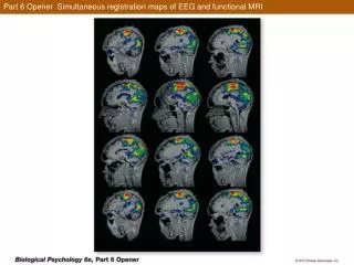
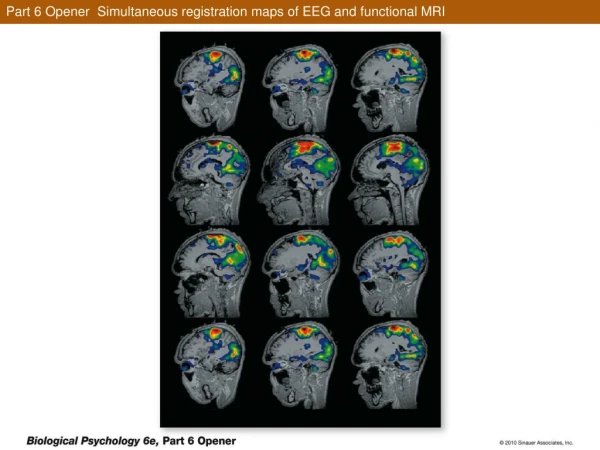
Part 6 Opener Simultaneous registration maps of EEG and functional MRI
Figure 17.1 Brain Tissue Removed from Henry Molaison (Patient H.M.)
Figure 17.3 Two Main Kinds of Memory: Declarative and Nondeclarative
Figure 17.3 Two Main Kinds of Memory: Declarative and Nondeclarative
Box 17.1 Learning and Memory: Some Basic Concepts and Definitions
Figure 17.5 Subtypes of Declarative and Nondeclarative Memory
Figure 17.6 Serial Position Curves from Immediate-Recall Experiments
Figure 17.7 Hypothesized Memory Processes: Encoding, Consolidation, and Retrieval
Figure 17.8 Encoding, Consolidation, and Retrieval of Declarative Memories
Figure 17.8 Encoding, Consolidation, and Retrieval of Declarative Memories
Figure 17.10 Memory Performance after Medial Temporal Lobe Lesions
Figure 17.10 Memory Performance after Medial Temporal Lobe Lesions
Figure 17.14 Tests of Specific Attributes of Memory (Part 1)
Figure 17.14 Tests of Specific Attributes of Memory (Part 2)
Figure 17.14 Tests of Specific Attributes of Memory (Part 3)
Figure 17.15 Brain Regions Involved in Different Kinds of Learning and Memory
Figure 17.15 Brain Regions Involved in Different Kinds of Learning and Memory (Part 1)
Figure 17.15 Brain Regions Involved in Different Kinds of Learning and Memory (Part 2)
Figure 17.16 Synaptic Changes That May Store Memories (Part 1)
Figure 17.16 Synaptic Changes That May Store Memories (Part 2)
Figure 17.17 Experimental Environments to Test the Effects of Enrichment on Learning and Brain Measures
Figure 17.20 Synaptic Plasticity Underlying Habituation in Aplysia
Figure 17.20 Synaptic Plasticity Underlying Habituation in Aplysia (Part 1)
Figure 17.20 Synaptic Plasticity Underlying Habituation in Aplysia (Part 2)
Figure 17.21 Long-Term Potentiation Occurs in the Hippocampus
Figure 17.21 Long-Term Potentiation Occurs in the Hippocampus (Part 1)
Figure 17.21 Long-Term Potentiation Occurs in the Hippocampus (Part 2)
Figure 17.21 Long-Term Potentiation Occurs in the Hippocampus (Part 3)
Figure 17.22 Roles of NMDA and AMPA Receptors in Induction of LTP in the CA1 Region
Figure 17.22 Roles of NMDA and AMPA Receptors in Induction of LTP in the CA1 Region (Part 1)
Figure 17.22 Roles of NMDA and AMPA Receptors in Induction of LTP in the CA1 Region (Part 2)
Figure 17.22 Roles of NMDA and AMPA Receptors in Induction of LTP in the CA1 Region (Part 3)
Figure 17.23 Steps in the Neurochemical Cascade during the Induction of LTP
Figure 17.23 Steps in the Neurochemical Cascade during the Induction of LTP
Figure 17.24 Functioning of the Neural Circuit for Conditioning of the Eye-Blink Reflex
Figure 17.24 Functioning of the Neural Circuit for Conditioning of the Eye-Blink Reflex (Part 1)