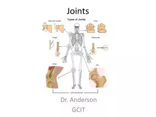
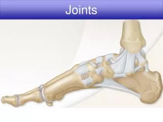
Joints
Joints. (c) Gomphosis. (a) Suture. (b) Syndesmosis. Joint held together with very short, interconnecting fibers, and bone edges interlock. Found only in the skull. Joint held together by a ligament. Fibrous tissue can vary in length, but is longer than in sutures.

Joints
E N D
Presentation Transcript
(c) Gomphosis (a) Suture (b) Syndesmosis Joint held together with very short, interconnecting fibers, and bone edges interlock. Found only in the skull. Joint held together by a ligament. Fibrous tissue can vary in length, but is longer than in sutures. “Peg in socket” fibrous joint. Periodontal ligament holds tooth in socket. Socket of alveolar process Suture line Fibula Tibia Root of tooth Dense fibrous connective tissue Ligament Periodontal ligament Figure 8.1 Fibrous joints.
(a) Suture Joint held together with very short, interconnecting fibers, and bone edges interlock. Found only in the skull. Suture line Dense fibrous connective tissue Figure 8.1a Fibrous joints.
(b) Syndesmosis Joint held together by a ligament. Fibrous tissue can vary in length, but is longer than in sutures. Fibula Tibia Ligament Figure 8.1b Fibrous joints.
(c) Gomphosis “Peg in socket” fibrous joint. Periodontal ligament holds tooth in socket. Socket of alveolar process Root of tooth Periodontal ligament Figure 8.1c Fibrous joints.
(a) Synchondroses Bones united by hyaline cartilage Sternum (manubrium) Epiphyseal plate (temporary hyaline cartilage joint) Joint between first rib and sternum (immovable) (b) Symphyses Bones united by fibrocartilage Body of vertebra Fibrocartilaginous intervertebral disc Hyaline cartilage Pubic symphysis Figure 8.2 Cartilaginous joints.
(a) Synchondroses Bones united by hyaline cartilage Sternum (manubrium) Epiphyseal plate (temporary hyaline cartilage joint) Joint between first rib and sternum (immovable) Figure 8.2a Cartilaginous joints.
(b) Symphyses Bones united by fibrocartilage Body of vertebra Fibrocartilaginous intervertebral disc Hyaline cartilage Pubic symphysis Figure 8.2b Cartilaginous joints.
Ligament Joint cavity (contains synovial fluid) Articular (hyaline) cartilage Fibrous capsule Articular capsule Synovial membrane Periosteum Figure 8.3 General structure of a synovial joint.
Tendon of quadriceps femoris Femur Suprapatellar bursa Articular capsule Patella Posterior cruciate ligament Subcutaneous prepatellar bursa Synovial cavity Lateral meniscus Lateral meniscus Infrapatellar fat pad Anterior cruciate ligament Deep infrapatellar bursa Tibia Patellar ligament (a) Sagittal section through the right knee joint Figure 8.8a The knee joint.
Anterior Anterior cruciate ligament Articular cartilage on lateral tibial condyle Articular cartilage on medial tibial condyle Lateral meniscus Medial meniscus Posterior cruciate ligament (b) Superior view of the right tibia in the knee joint, showing the menisci and cruciate ligaments Figure 8.8b The knee joint.
Quadriceps femoris muscle Tendon of quadriceps femoris muscle Patella Medial patellar retinaculum Lateral patellar retinaculum Tibial collateral ligament Fibular collateral ligament Patellar ligament Tibia Fibula (c) Anterior view of right knee Figure 8.8c The knee joint.
Femur Tendon of adductor magnus Articular capsule Oblique popliteal ligament Medial head of gastrocnemius muscle Lateral head of gastrocnemius muscle Popliteus muscle (cut) Bursa Fibular collateral ligament Tibial collateral ligament Arcuate popliteal ligament Tendon of semimembranosus muscle Tibia (d) Posterior view of the joint capsule,including ligaments Figure 8.8d The knee joint.
Posterior cruciate ligament Fibular collateral ligament Medial condyle Tibial collateral ligament Lateral condyle of femur Anterior cruciate ligament Lateral meniscus Medial meniscus Tibia Patellar ligament Patella Fibula Quadriceps tendon (e) Anterior view of flexed knee, showing the cruciateligaments (articular capsule removed, and quadricepstendon cut and reflected distally) Figure 8.8e The knee joint.
Medial femoral condyle Anterior cruciate ligament Medial meniscus on medial tibial condyle Patella (f) Photograph of an opened knee joint; view similar to (e) Figure 8.8f The knee joint.
Acromion of scapula Coracoacromial ligament Joint cavity containing synovial fluid Subacromial bursa Fibrous articular capsule Hyaline cartilage Tendon sheath Synovial membrane Tendon of long head of biceps brachii muscle Fibrous capsule Humerus (a) Frontal section through the right shoulder joint Figure 8.4a Bursae and tendon sheaths.
Coracoacromial ligament Subacromial bursa Humerus resting Cavity in bursa containing synovial fluid Bursa rolls and lessens friction. Humerus head rolls medially as arm abducts. Humerus moving (b) Enlargement of (a), showing how a bursaeliminates friction where a ligament (or otherstructure) would rub against a bone Figure 8.4b Bursae and tendon sheaths.
f Nonaxial Uniaxial Biaxial Multiaxial c b Plane joint (intercarpal joint) a a e d Figure 8.7a Types of synovial joints.
f Nonaxial Uniaxial Biaxial Multiaxial c b Hinge joint (elbow joint) b a e d Figure 8.7b Types of synovial joints.
f Nonaxial Uniaxial Biaxial Multiaxial c b c Pivot joint (proximal radioulnar joint) a e d Figure 8.7c Types of synovial joints.
f Nonaxial Uniaxial Biaxial Multiaxial c b d Condyloid joint (metacarpophalangeal joint) a e d Figure 8.7d Types of synovial joints.
f Nonaxial Uniaxial Biaxial Multiaxial c b e Saddle joint (carpometacarpal joint of thumb) a e d Figure 8.7e Types of synovial joints.
f Nonaxial Uniaxial Biaxial Multiaxial c b f Ball-and-socket joint (shoulder joint) a e d Figure 8.7f Types of synovial joints.
Lateral Medial Patella (outline) Hockey puck Tibial collateral ligament (torn) Medial meniscus (torn) Anterior cruciate ligament (torn) Figure 8.9 A common knee injury.
Torn meniscus Figure 8.14 Arthroscopic photograph of a torn medial meniscus.
Figure 8.15 X ray of a hand deformed by rheumatoid arthritis.
Table 8.2 Structural and Functional Characteristics of Body Joints (1 of 4)
Table 8.2 Structural and Functional Characteristics of Body Joints (2 of 4)
Table 8.2 Structural and Functional Characteristics of Body Joints (3 of 4)
Table 8.2 Structural and Functional Characteristics of Body Joints (4 of 4)
A Closer Look 8.1a Joints: From Knights in Shining Armor to Bionic Humans
A Closer Look 8.1b: Joints: From Knights in Shining Armor to Bionic Humans