Principles of Protein Structure
470 likes | 854 Vues
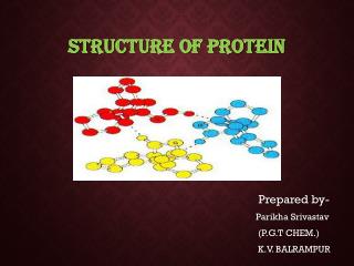
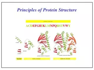
Principles of Protein Structure. primary structure. ACD EFGH I K LM NPQ RST VW Y. NH 2 Lys ine His tidine Val ine Arg inine Ala nine COOH. Different Levels of Protein Structure. Common Secondary Structure Elements. The Alpha Helix. Properties of alpha helix.

Principles of Protein Structure
E N D
Presentation Transcript
Principles of Protein Structure primary structure ACDEFGHIKLMNPQRSTVWY
NH2 Lysine Histidine Valine Arginine Alanine COOH Different Levels of Protein Structure
Common Secondary Structure Elements The Alpha Helix
Properties of alpha helix • 3.6 residues per turn, 13 atoms between H-bond donor and acceptor • approx. -60º; approx. -40º • H- bond between C=O of ith residue & -NH of (i+4)th residue • First -NH and last C=O groups at the ends of helices do not participate in H-bond • Ends of helices are polar, and almost always at surfaces of proteins • Always right- handed • Macro- dipole
Helical wheel Residues i, i+4, i+7 occur on one face of helices, and hence show definite pattern of hydrophobicity/ hydrophilicity
Association of helices: coiled coils Introduction to Molecular Biophysics These coiled coils have a heptad repeat abcdefg with nonpolar residues at position a and d and an electrostatic interaction between residues e and g. Isolated alpha helices are unstable in solution but are very stable in coiled coil structures because of the interactions between them The chains in a coiled-coil have the polypeptide chains aligned parallel and in exact axial register. This maximizes coil formation between chains. The coiled coil is a protein motif that is often used to control oligomerization. They involve a number of alpha-helices wound around each other in a highly organised manner, similar to the strands of a rope.
The Leucine Zipper Coiled Coil Introduction to Molecular Biophysics Initially identified as a structural motif in proteins involved in eukaryotic transcription. (Landschultz et al., Science 240: 1759-1763 (1988). Originally identified in the liver transcription factor C/EBP which has a Leu at every seventh position in a 28 residue segment.
Association of helices: coiled coils The helices do not have to run in the same direction for this type of interaction to occur, although parallel conformation is more common. Antiparallel conformation is very rare in trimers and unknown in pentamers, but more common in intramolecular dimers, where the two helices are often connected by a short loop. Chan et al., Cell 89, Pages 263-273.
Basis for the helical dipole In an alpha helix all of the peptide dipoles are oriented along the same direction. Consequently, the alpha helix has a net dipole moment. • Since the dipole moment of a peptide bond is 3.5 Debye units, the alpha • helix has a net macrodipole of: • n X 3.5 Debye units (where n= number of residues) • This is equivalent to 0.5 – 0.7 unit charge at the end of the helix. • The amino terminus of an alpha helix is positive and the • carboxy terminus is negative.
Structure of human TIM • Two helix dipoles are seen to play important roles: • Stabilization of inhibitor 2-PG • Modulation of pKa of active site His-95.
Helical Propensities Ala -0.77 Arg -0.68 Lys -0.65 Leu -0.62 Met -0.50 Trp -0.45 Phe -0.41 Ser -0.35 Gln -0.33 Glu -0.27 Cys -0.23 Ile -0.23 Tyr -0.17 Asp -0.15 Val -0.14 Thr -0.11 Asn -0.07 His -0.06 Gly 0 Pro ~3
Common Secondary Structure Elements The Beta Sheet
Secondary Structure:Phi & Psi Angles Defined • Rotational constraints emerge from interactions with bulky groups (ie. side chains). • Phi & Psi angles define the secondary structure adopted by a protein.
The dihedral angles at Ca atom of every residue provide polypeptides requisite conformational diversity, whereby the polypeptide chain can fold into a globular shape
Table 10 Secondary Structure
Beyond Secondary Structure • Supersecondary structure (motifs): small, discrete, commonly observed aggregates of secondary structures • b sheet • helix-loop-helix • bab • Domains: independent units of structure • b barrel • four-helix bundle • *Domains and motifs sometimes interchanged*
Supersecondary structure: Crossovers in b-a-b-motifs Left handed Right handed
EF Hand • Consists of two perpendicular 10 to 12 residue alpha helices with a 12-residue loop region between • Form a single calcium-binding site (helix-loop-helix). • Calcium ions interact with residues contained within the loop region. • Each of the 12 residues in the loop region is important for calcium coordination. • In most EF-hand proteins the residue at position 12 is a glutamate. The glutamate contributes both its side-chain oxygens for calcium coordination. Calmodulin, recoverin : Regulatory proteins Calbindin, parvalbumin: Structural proteins
EF Fold Found in Calcium binding proteins such as Calmodulin
Helix Turn Helix Motif • Consists of two a helices and a short extended amino acid chain between them. • Carboxyl-terminal helix fits into the major groove of DNA. • This motif is found in DNA-binding proteins, including l repressor, tryptophan repressor, catabolite activator protein (CAP)
Rossman Fold • The beta-alpha-beta-alpha-beta subunit • Often present in nucleotide-binding proteins
What is a Protein Fold? • Compact, globular folding arrangement of the polypeptide chain • Chain folds to optimise packing of the hydrophobic residues in the interior core of the protein
Tertiary structure examples: All-a Cytochrome Cfour-helix bundle AlamethicinThe lone helix Rophelix-turn-helix
Tertiary structure examples: All-b b sandwich b barrel
Tertiary structure examples: a/b placental ribonucleaseinhibitor a/b horseshoe triose phosphateisomerase a/b barrel
Four helix bundle • 24 amino acid peptide with a hydrophobic surface • Assembles into 4 helix bundle through hydrophobic regions • Maintains solubility of membrane proteins
TIM Barrel • The eight-stranded a /b barrel (TIM barrel) • The most common tertiary fold observed in high resolution protein crystal structures • 10% of all known enzymes have this domain
Domains are independently folding structural units. Often, but not necessarily, they are contiguous on the peptide chain. Often domain boundaries are also intron boundaries.
Domain swapping: Parts of a peptide chain can reach into neighboring structural elements: helices/strands in other domains or whole domains in other subunits. Domain swapped diphteria toxin:
Transmembrane Motifs • Helix bundlesLong stretches of apolar amino acidsFold into transmembrane alpha-helices“Positive-inside rule”Cell surface receptors Ion channels Active and passive transporters • Beta-barrelAnti-parallel sheets rolled into cylinderOuter membrane of Gram-negative bacteria Porins (passive, selective diffusion)
Quaternary Structure • Refers to the organization of subunits in a protein with multiple subunits • Subunits may be identical or different • Subunits have a defined stoichiometry and arrangement • Subunits held together by weak, noncovalent interactions (hydrophobic, electrostatic) • Associate to form dimers, trimers, tetramers etc. (oligomer) • Typical Kd for two subunits: 10-8 to 10-16M (tight association) • Entropy loss due to association - unfavorable • Entropy gain due to burying of hydrophobic groups - very favourable
Stability: reduction of surface to volume ratio Genetic economy and efficiency Bringing catalytic sites together Cooperativity (allostery) Structural and functional advantages of quaternary structure