Download
1 / 27
270 likes | 481 Vues
Scanning Microscope for muon radiography with nuclear emulsion.

E N D
Scanning Microscopefor muon radiographywith nuclear emulsion Cristiano Bozza1, Lucia Consiglio2, Nicola D'Ambrosio3, Giovanni De Lellis4, Chiara De Sio5, Natalia Di Marco3, UmutKose6, Eduardo Medinaceli7, Seigo Miyamoto8, RyuichiNishiyama8, Fabio Pupilli3, Simona Maria Stellacci1, Chiara Sirignano7, Paolo Strolin4, Hiroyuki Tanaka8, Valeri Tioukov2 University of Salerno and INFN1, INFN Napoli2, INFN / LNGS3, University of Napoli and INFN4,Universityof Salerno5,INFN Padova6,University of Padova and INFN7,Universityof Tokyo8
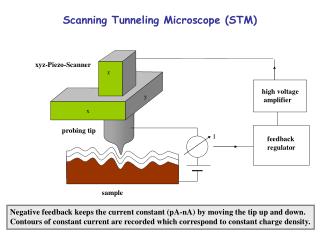
Scanning Microscope for muon radiography with nuclear emulsion Nuclearemulsiondetectorsformuonradiography Detectors are made of stacked emulsion films m m e+e- e+e- e+e- Emulsion has no time resolution, no trigger: all tracks are recorded Emulsion films record hard tracks as well as soft tracks 3D information available for each track: momentum discrimination and/or particle id. possible!
Scanning Microscope for muon radiography with nuclear emulsion Nuclearemulsionimages AgBr gel 1 μm ChargedparticlesionizeAgatoms (stochasticprocess), producing the latentimage Metallic Ag grows in filaments during development With green-white light the average l is 600 nm: the filaments cannot be resolved because of diffraction “Grains” = clusters of filaments
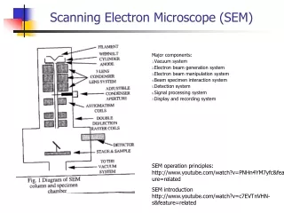
Scanning Microscope for muon radiography with nuclear emulsion Looking at emulsionfilms: basicopticalsetup CMOS sensor Objective lens (or lens system) Illuminated spot Emulsion film Plastic base Condenser lens Lamp (optionally w/ filters) White, green or blue
Scanning Microscope for muon radiography with nuclear emulsion Nuclearemulsionimages Imaging by objective + camera: the spatial density of metallic Ag is folded with the PSF (point-spread function), characterizing the optical setup Out of focus Y(x,y,z) Focal plane Out of focus Depthoffield: ~3 μm Typical grain size after development: 0.2÷1 μm (0.5 μm in the case shown in this talk) 50 μm Grains in emulsion image: high-energy tracks, electrons, fog (randomly developed grains,not touched by any ionizing particle)
Scanning Microscope for muon radiography with nuclear emulsion Nuclearemulsionimages Grain images are not uniform, and depend more on the neighborhoods than on thefeatures of grains themselves Finite depth of field: grains out of focus can be seen Shadow effect: grains stacked one on top of the other on the focal axis look darker (bigger) Highly ionizing particles: grains may be so close they cannot be resolved A “hole”: no doubt the charged particle passed there, but it just did not ionize!
Scanning Microscope for muon radiography with nuclear emulsion Nuclearemulsionimages 3D tomography:change focal plane Alignment residuals of track grains: 50 nm in optical microscopy! Good precision helps rejecting random alignments and thus increasing signal/background ratio
Scanning Microscope for muon radiography with nuclear emulsion The European Scanning System (ESS) Developed for OPERA, used in all European laboratories Also installed at Tokyo ERI Scanning speed: 20 cm2/h/side Z stage (Micos)0.05 μm nominalprecision CMOS camera1280×1024 pixel256 graylevels376 frames/sec(Mikrotron MC1310) Emulsion Plate XY stage (Micos)0.1 μm nominalprecision Illumination system, objective (Oil 50× NA 0.85) and optical tube (Nikon)
Scanning Microscope Camera Vision Processor(Matrox Odyssey) Host PC(Dual PentiumWorkstation)RunningWinXP 2D Images(peak 452 MB/s,avg. 97 MB/s) Binarized2D Images Motors(VEXTA Nanostep) Motion Controller(National InstrumentsFlexMotion) Grains XYZ MotionCommands Power TomographicsequencesZ axismoving, 2D imagesspanningemulsionthickness 280×365μm2 Next field of view,Z at top, new cycle Process/save data Move XYZ to next view for muon radiography with nuclear emulsion The ESS: workingprinciples DAQ cycle(185 ms) Functional blocks
Scanning Microscope for muon radiography with nuclear emulsion The ESS: Image processing (SySal2000) 15 images10 grainssignal/image,3000 grainsbackground+noise, shadows, scratches, spots 2D FIR Filter+Equalization+ Threshold Grainrecognition(Host PC, multithreadedAssembler code) 3D microtrackreconstruction(HostPC or trackingservers, multithreadedC++ code) 300 ÷ 3000 microtracks / view
Scanning Microscope for muon radiography with nuclear emulsion The ESS: Tracking(SySal2000) Tracking: recognitionofalignedsequencesofgrains in 2D images(microtracks) Highlyoptimizedalgorithmtodeal with big combinatorialcomplexity Parallel processing: can use upto 8 processors/cores per machine
Scanning Microscope for muon radiography with nuclear emulsion The ESS: Tracking(SySal2000) ScanGrid: usepowerfulmachinesdedicatedto on-line tracking/computing and simplify the architectureofdata-taking Grains Other info (setup, monitoring) tracks Installation in Salerno: 70 trackingcoressharedby 4 microscopes Installation at LNGS: 80 trackingcoressharedby 10 microscopes Automaticloadbalancing (differentqualityofemulsionrequiresdifferent processing power)
Scanning Microscope for muon radiography with nuclear emulsion The ESS: currentperformances Tests on 8 GeV/c pionbeams Microtrack Base-track Sy = 0 Sy = -0.180 Notice: efficiencydepends on emulsionquality!!!
Scanning Microscope for muon radiography with nuclear emulsion The ESS: currentperformances Precisionoffilm-to-filmtrack connection Sx = 0.025Sy = 0 Sx = 0.600Sy = -0.180
Scanning Microscope for muon radiography with nuclear emulsion The ESS: currentperformances Precisionoffilm-to-filmtrack connection Sx = 0.025Sy = 0 Sx = 0.600Sy = -0.180
Scanning Microscope for muon radiography with nuclear emulsion The Quick Scanning System (QSS): evolutionof the ESS • Increase scanning speed: enable using larger areas higher statistics • improve signal/background ratio • improve sensitivity to flux variations • improve sensitivity to density variations • Increase sensor size (area scanned at each tomographic sweep) • Increase grabbing speed • Increase processing speed • Reduce dead time due to motion Keep data quality high: low number of fake tracks effectively discriminate electrons from muons good precision effectively discriminate electrons from muons high efficiency increase data rate/unit area(with triplet or quadruplet stacks, microtracking efficiency suppresses statistics with at least the 6th power!)
Scanning Microscope for muon radiography with nuclear emulsion The QSS Same mechanics, new hardware Double CL frame grabber (MatroxRadient) New optics (20×) 4 Mpixel camera, 400 fps Image processing and tracking by GPU New motion control unit Pro-DexMAXk
Scanning Microscope for muon radiography with nuclear emulsion The Quick Scanning System (QSS): evolutionof the ESS Z motion, 2D imagesread and processed on-the-fly (vision processor in host PC) “Stop’n’go” “Continuous motion”X axis travels at constant speed 2D images read and processed on-the-fly (GPU in host PC) Dead time due to motion is reduced
Scanning Microscope for muon radiography with nuclear emulsion The Quick Scanning System (QSS): evolutionof the ESS Stepsfortrackreconstruction Dark pixel clustering GPU View-to-viewalignmentbychain pattern matching GPU Image-to-imagealignment (sameview) GPU Microtrack recognition GPU Chainsofclusters (1 chain = 1 or more grains) GPU Base-tracklinking (uses standard SySal.NET linking) CPU
Scanning Microscope for muon radiography with nuclear emulsion The Quick Scanning System (QSS): evolutionof the ESS • Image-to-imagealignment • 1 ms stage samplingloop on XYZ (doesnotrequirepiezodrive) • Imagedistortioncorrections XY curvature Z curvature Z axisslant(X and Y) XY Trapezium Magnificationvs. Z
Scanning Microscope for muon radiography with nuclear emulsion The Quick Scanning System (QSS): evolutionof the ESS Image-to-imagealignmentresults mm mm XY precision: 0.12 mm
Scanning Microscope for muon radiography with nuclear emulsion The Quick Scanning System (QSS): evolutionof the ESS View-to-view mapping:discard duplicated grains in overlapregion and correct misalignmentsdue to stage motion 3D track recognition: Microtracks: sequences of aligned grains (230 nm tolerance on X/Y, 2 μm on Z)recognized in the whole scanning volume GPU-based tracking server (2× NVidia GTX690) – 3072 CUDA cores/microscope
Scanning Microscope for muon radiography with nuclear emulsion The Quick Scanning System (QSS): evolutionof the ESS • View-to-view 3D pattern matching • Recoverstransversevibrations and XYZ samplingerrors (allowsmicrotrackingacrossviews) • Mergeschainduplicates in overlap volume (preventsexcess microtracks) mm mm Z precision: 2.6 mm mm XY precision: 0.2 mm
Scanning Microscope for muon radiography with nuclear emulsion The Quick Scanning System (QSS): evolutionof the ESS Tracking on test filmsexposedtolarge angle beams Preliminary!
Scanning Microscope for muon radiography with nuclear emulsion The Quick Scanning System (QSS): evolutionof the ESS Tracking on test filmsexposedtolarge angle beams Preliminary!
Scanning Microscope for muon radiography with nuclear emulsion The Quick Scanning System (QSS): evolutionof the ESS The scanning speed is 41 cm2/h with 31 layers, 58% view travel, 200 fps operation This should ensure maximum signal/noise ratio Depending on conditions, it is possible to speed up the system with minimal changes (tuning quantities in the parameter form) 3121 layers (glued emulsions, low fog) 64 cm2/h (tested) + 58%75% view travel (8 core CPU) 85 cm2/h (technically tested) + 200400 fps 120 cm2/h (estimated) For future applications, having low fog emulsion will in general improve the performances (fewer layers needed for high efficiency, low combinatorial background) Cost of the upgrade from ESS to QSS: about 20 k€!
Scanning Microscope for muon radiography with nuclear emulsion Conclusions The ESS is an established technology that provides the performances needed for muon radiography Film-to-film connection at micrometric level (slope accuracy of the order of 10 mrad is easily achieved) The ESS has been used for Unzen and Stromboli data readout The QSS is the upgraded project that is approaching its first stage (2× speed) with cheap hardware upgrades Outlook: 6× speed increasing just the number of GPU’s (4 × 600 €) Stay tuned for first applications of the QSS in muon radiography!