Popliteal fossa
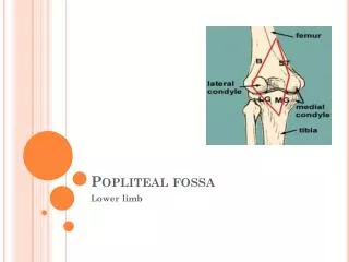
Popliteal fossa. Lower limb. Popliteal fossa. The popliteal fossa is a diamond-shaped intermuscular space situated at the back of the knee


Popliteal fossa
E N D
Presentation Transcript
Poplitealfossa Lower limb
Poplitealfossa • The poplitealfossa is a diamond-shapedintermuscular space situated at the back of the knee • The poplitealfossa is an important area of transition between the thigh and leg and is the major route by which structures pass from one region to the other.
Boundaries • Superolaterally :Biceps femoris • Superomedially:Semimembranosus, lateral to which is the semitendinosus • Inferolaterally and inferomedially: Lateral and medial heads of the gastrocnemius. • Posteriorly(roof): Skin and poplitealfascia • The anterior wall or floor: Poplitealsurface of the femur and the popliteusmuscle.
Contents of the poplitealfossa • 1. Termination of the small saphenous vein. • 2. Popliteal arteries and veins. • 3. Tibial and common fibular nerves. • 4. Posterior cutaneous nerve of thigh • 5. Popliteal lymph nodes and lymphatic vessels
Neurovascular Structures and Relationships in the PoplitealFossa • Posterior to anterior as in dissection : • 1. Nervesare encountered first • 2. Veins • 3. Arterieslie deepest, directly on the surface of the femur.
Blood Vessels in the PoplitealFossa • Popliteal artery: • Continuation of the femoral artery , begins when the latter passes through the adductor hiatus. • The popliteal artery is the deepest of the neurovascular structures in the poplitealfossa and is therefore difficult to palpate; however, a pulse can usually be detected by deep palpation medial to the midline. • The popliteal artery passes inferolaterally through the fossa and ends at the inferior border of the popliteus by dividing into the anterior and posteriortibial arteries.
popliteal artery • In the poplitealfossa, the popliteal artery gives rise to branches, which supply adjacent muscles, and to a series of geniculate arteries, which contribute to vascular anastomoses around the knee. • The genicular arteries are the superior lateral, superior medial, middle, inferior lateral, and inferior medial genicular arteries
Genicularanastomosis • 1. Descending genicularbranch of the femoral artery, superomedially. • 2. Descending branch of the lateral femoral circumflex artery, superolaterally. • 3. Anterior tibial recurrent branch of the anterior tibial artery, inferolaterally.
popliteal vein • The popliteal vein begins at the distal border of the popliteus as a continuation of the posterior tibial vein. • The small saphenous vein passes from the posterior aspect of the lateral malleolus to the poplitealfossa, where it pierces the deep popliteal fascia and enters the popliteal vein.
Nerves in the PoplitealFossa • The sciatic nerve usually ends at the superior angle of the poplitealfossa by dividing into the tibialand common fibular nerves.
tibial nerve • It runs downward through the poplitealfossa, lying first on the lateral side of the popliteal artery, then posterior to it, and finally medial to it. • Branches • 1. Cutaneous:Thesural nerve descends between the two heads of the gastrocnemius muscle and is usually joined by the sural communicating branch of the common peroneal nerve. • This nerve supplies the lateral side of the leg and ankle. • 2. Muscular: gastrocnemius and the plantaris, soleus, and popliteus
common fibular nerve • Branches • 1. Cutaneous: The sural communicating branch runs downward and joins the sural nerve. • 2. Muscular: branch to the short head of the biceps femoris muscle, which arises high up in the poplitealfossa
Lymph Nodes in the PoplitealFossa • Superficial popliteal lymph nodes • Deep popliteal lymph nodes