Practical Approach to Acute Gastrointestinal Bleeding
960 likes | 1.47k Vues
Practical Approach to Acute Gastrointestinal Bleeding. Christopher S. Huang MD Assistant Professor of Medicine Boston University School of Medicine Section of Gastroenterology Boston Medical Center. Learning Objectives. UGIB Nonvariceal (PUD) and variceal

Practical Approach to Acute Gastrointestinal Bleeding
E N D
Presentation Transcript
Practical Approach to Acute Gastrointestinal Bleeding Christopher S. Huang MD Assistant Professor of Medicine Boston University School of Medicine Section of Gastroenterology Boston Medical Center
Learning Objectives • UGIB • Nonvariceal (PUD) and variceal • Resuscitation, risk assessment, pre-endoscopy management • Role of endoscopy • Post-endoscopy management • LGIB • Risk assessment • Role and timing of colonoscopy • Non-endoscopic diagnostic and treatment options
Definitions • Upper GI bleed – arising from the esophagus, stomach, or proximal duodenum • Mid-intestinal bleed – arising from distal duodenum to ileocecal valve • Lower intestinal bleed – arising from colon/rectum
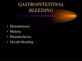
Stool color and origin/pace of bleeding • Guaiac positive stool • Occult blood in stool • Does not provide any localizing information • Indicates slow pace, usually low volume bleeding • Melena • Very dark, tarry, pungent stool • Usually suggestive of UGI origin (but can be small intestinal, proximal colon origin if slow pace) • Hematochezia • Spectrum: bright red blood, dark red, maroon • Usually suggestive of colonic origin (but can be UGI origin if brisk pace/large volume)
Case Vignette – CC: • 68 yo male presents with a chief complaint of a large amount of “bleeding from the rectum”
Case Vignette - HPI • Describes bleeding as large volume, very dark maroon colored stool • Has occurred 4 times over past 3 hours • He felt light headed and nearly passed out upon trying to get up to go the bathroom
Case Vignette - HPI • Denies abdominal pain, nausea, vomiting, antecedent retching • No history of heartburn, dysphagia, weight loss • No history of diarrhea or constipation/hard stools • No prior history of GIB • Screening colonoscopy 10 years ago – no polyps, (+) diverticulosis
Case Vignette – PMHx, Meds • Hepatitis C • CAD – h/o MI • PVD • AAA – s/p elective repair 3 years ago • HTN • Hypercholesterolemia • Lumbago • Medications: • Aspirin • Clopidogrel • Atorvastatin • Atenolol • Lisinopril
Case Vignette – Physical Exam • Physical examination: • BP 105/70, Pulse 100, (+) orthostatic changes • Alert and mentating, but anxious appearing • Anicteric • Mid line scar, benign abdomen, nontender liver edge palpable in epigastrium, no splenomegaly • Rectal examination – no masses, dark maroon blood
Case Vignette - Labs • Labs • Hct 21% (Baseline 33%) • Plt 110K • BUN 34, Cr 1.0 • Alb 3.5 • INR 1.6 • ALT 51, AST 76
Initial Considerations • Differential diagnosis? • What is most likely source? • What diagnosis can you least afford to miss? • How sick is this patient? (risk stratification) • Determines disposition • Guides resuscitation • Guides decision re: need for/timing of endoscopy
Differential Diagnosis – Upper GIB • Peptic ulcer disease • Gastroesophageal varices • Erosive esophagitis/gastritis/duodenitis • Mallory Weiss tear • Vascular ectasia • Neoplasm • Dieulafoy’s lesion • Aortoenteric fistula • Hemobilia, hemosuccus pancreaticus Most common Rare, but cannot afford to miss
Differential Diagnosis – Lower GIB Most common diagnosis • Diverticulosis • Angioectasias • Hemorrhoids • Colitis (IBD, Infectious, Ischemic) • Neoplasm • Post-polypectomy bleed (up to 2 weeks after procedure) • Dieulafoy’s lesion
History and Physical History Physical Examination Vital signs, orthostatics Abdominal tenderness Skin, oral examination Stigmata of liver disease Rectal examination Objective description of stool/blood Assess for mass, hemorrhoids No need for guaiac test • Localizing symptoms • History of prior GIB • NSAID/aspirin use • Liver disease/cirrhosis • Vascular disease • Aortic valvular disease, chronic renal failure • AAA repair • Radiation exposure • Family history of GIB
History and Physical History Physical Examination Vital signs, orthostatics Abdominal tenderness Skin, oral examination Stigmata of liver disease Rectal examination Objective description of stool/blood Assess for mass, hemorrhoids No need for guaiac test • Localizing symptoms • History of prior GIB • NSAID/aspirin use • Liver disease/cirrhosis • Vascular disease • Aortic valvular disease, chronic renal failure • AAA repair • Radiation exposure • Family history of GIB
Take Home Point # 1 • Always get objective description of stool • Avoid noninformative terms such as “grossly guaiac positive”
Take Home Point #2 • If you need a card to tell you whether there’s blood in the stool, it’s NOT an acute GIB
Narrowing the DDx: Upper or Lower Source? • Predictors of UGI source: • Age <50 • Melenic stool • BUN/Creatinine ratio • If ratio ≥ 30, think upper GIB J ClinGastroenterol 1990;12:500 Am J Gastroenterol 1997;92:1796 Am J Emerg Med 2006;24:280
Utility of NG Tube • Most useful situation: patients with severe hematochezia, and unsure if UGIB vs. LGIB • Positive aspirate (blood/coffee grounds) indicates UGIB • Can provide prognostic info: • Red blood per NGT – predictive of high risk endoscopic lesion • Coffee grounds – less severe/inactive bleeding • Negative aspirate – not as helpful; 15-20% of patients with UGIB have negative NG aspirate Ann Emerg Med 2004;43:525 Arch Intern Med 1990;150:1381 GastrointestEndosc 2004;59:172
Take Home Point #3 Upper GI bleed must still be considered in patients with severe hematochezia, even if NG aspirate negative
Initial Assessment • Always remember to assess A,B,C’s • Assess degree of hypovolemic shock
Resuscitation • IV access: large bore peripheral IVs best (alt: cordis catheter) • Use crystalloids first • Anticipate need for blood transfusion • Threshold should be based on underlying condition, hemodynamic status, markers of tissue hypoxia • Should be administered if Hgb ≤ 7 g/dL • 1 U PRBC should raise Hgb by 1 (HCT by 3%) • Remember that initial Hct can be misleading (Hct remains the same with loss of whole blood, until re-equilibration occurs) • Correct coagulopathy
Resuscitation • IV access: large bore peripheral IVs best (alt: cordis catheter) • Use crystalloids first • Anticipate need for blood transfusion • Threshold should be based on underlying condition, hemodynamic status, markers of tissue hypoxia • Should be administered if Hgb ≤ 7 g/dL • 1 U PRBC should raise Hgb by 1 (HCT by 3%) • Remember that initial Hct can be misleading (Hct remains the same with loss of whole blood, until re-equilibration occurs) • Correct coagulopathy 40% 40% bleed Time IVFs 20%
Transfusion Strategy • Randomized trial: • 921 subjects with severe acute UGIB • Restrictive (tx when Hgb<7; target 7-9) vs. Liberal (tx when Hgb<9; target 9-11) • Primary outcome: all cause mortality rate within 45 days NEJM 2013;368;11-21
Restrictive Strategy Superior Benefit seen primarily in Child A/B cirrhotics NEJM 2013;368;11-21
Resuscitation • IV access: large bore peripheral IVs best (alt: cordis catheter) • Use crystalloids first • Anticipate need for blood transfusion • Threshold should be based on underlying condition, hemodynamic status, markers of tissue hypoxia • Should be administered if Hgb ≤ 7 g/dL • 1 U PRBC should raise Hgb by 1 (HCT by 3%) • Remember that initial Hct can be misleading (Hct remains the same with loss of whole blood, until re-equilibration occurs) • Correct coagulopathy Weigh risks and benefits of reversing anticoagulation Assess degree of coagulopathy Vitamin K – slow acting, long-lived FFP – fast acting, short lived - Give 1 U FFP for every 4 U PRBCs
Resuscitation • Early intensive resuscitation reduces mortality • Consecutive series of patients with hemodynamically significant UGIB • First 36 subjects = Observation Group (no intervention) • Second 36 subjects = Intensive Resuscitation Group (intense guidance provided) – goal was to decrease time to correction of hemodynamics, Hct and coagulopathy Am J Gastroenterol 2004;99:619
Early Intensive Resuscitation Reduces UGIB Mortality Intervention: Faster correction of hemodynamics, Hct and coags. Time to endoscopy similar (groups are essentially the same) Am J Gastroenterol 2004;99:619
Early Intensive Resuscitation Reduces UGIB Mortality • Observation group • 5 MI • 4 deaths • Intense group • 2 MI • 1 death (sepsis) Am J Gastroenterol 2004;99:619
Causes of Mortality in Patients with Peptic Ulcer Bleeding • Patients rarely bleed to death • Prospective cohort study >10,000 cases of peptic ulcer bleed • Mortality rate 6.2% • 80% of deaths not related to bleeding Am J Gastroenterol 2010;105:84
Causes of Mortality in Patients with Peptic Ulcer Bleeding • Most common causes of non-bleeding mortality: • Terminal malignancy (34%) • Multiorgan failure (24%) • Pulmonary disease (24%) • Cardiac disease (14%) Am J Gastroenterol 2010;105:84
Take Home Point #4 Early resuscitation and supportive measures are critical to reduce mortality from UGIB
Risk Stratification • Identify patients at high risk for adverse outcomes • Helps determine disposition (ICU vs. floor vs. outpatient) • May help guide appropriate timing of endoscopy
Rockall Scoring System • Validated predictor of mortality in patients with UGIB • 2 components: clinical + endoscopic Gut 1996;38:316
AIMS65 • Simple risk score that predicts in-hospital mortality, LOS, cost in patients with acute UGIB Albumin <3.0 INR > 1.5 Mental status altered Systolic BP <90 65+ years old GastrointestEndosc 2011;74:1215
AIMS65 GastrointestEndosc 2011;74:1215
Blatchford Score • Predicts need for endoscopic therapy • Based on readily available clinical and lab data • Can use UpToDate calculator Lancet 2000;356:1318
Blatchford Score GastrointestEndosc 2010;71:1134
Blatchford Score • Most useful for safely discriminating low risk UGIB patients who will likely NOT require endoscopic hemostasis • “Fast track Blatchford” – patient at low risk if: BUN < 18 mg/dL Hgb > 13 (men), 12 (women) SBP >100 HR < 100
Pre-endoscopic Pharmacotherapy • For Non-Variceal UGIB • IV PPI: 80 mg bolus, 8 mg/hr drip • Rationale: suppress acid, facilitate clot formation and stabilization • Duration: at least until EGD, then based on findings
Pre-endoscopy PPI • Reduces the proportion of patients with high risk endoscopic stigmata (“downstages” lesion) • Decreases need for endoscopic therapy • Has not been shown to reduce rebleeding, surgery, or mortality rates High risk Low risk Endoscopic treatment required: Omeprazole – 19% (23% of PUD) Placebo – 28% (37% of PUD) N Engl J Med 2007;356:1631
Endoscopy - Nonvariceal UGIB • Early endoscopy (within 24 hours) is recommended for most patients with acute UGIB • Achieves prompt diagnosis, provides risk stratification and hemostasis therapy in high-risk patients J ClinGastroenterol 1996;22:267 GastrointestEndosc 1999;49:145 Ann Intern Med 2010;152:101
When is Endoscopic Therapy Required? • ~80% bleeds spontaneously resolve • Endoscopic stigmata of recent hemorrhage major
Major Stigmata – Active Spurting Kelsey, PB (Dec 04 2003). Duodenum - Ulcer, Arterial Spurting, Treated with Injection and Clip. The DAVE Project. Retrieved Aug, 1, 2010, from http://daveproject.org/viewfilms.cfm?film_id=39
Adherent Clot • Role of endoscopic therapy of ulcers with adherent clot is controversial • Clot removal usually attempted • Underlying lesion can then be assessed, treated if necessary
Minor Stigmata Flat pigmented spot Clean base Low rebleeding risk – no endoscopic therapy needed
Endoscopic Hemostasis Therapy • Epinephrine injection • Thermal electrocoagulation • Mechanical (hemoclips) • Combination therapy superior to monotherapy Kelsey, PB (Nov 08 2005). Stomach - Gastric Ulcer, Visible Vessel. The DAVE Project. Retrieved Aug, 1, 2010, from http://daveproject.org/viewfilms.cfm?film_id=306 Baron, TH (May 01 2007). Duodenum - Bleeding Ulcer Treated with Thermal Therapy, Perforation Closed with Hemoclips. The DAVE Project. Retrieved Aug, 1, 2010, from http://daveproject.org/viewfilms.cfm?film_id=620
Nonvariceal UGIB –Post-endoscopy management • Patients with low risk ulcers can be fed promptly, put on oral PPI therapy. • Patients with ulcers requiring endoscopic therapy should receive PPI gtt x 72 hours • Significantly reduces 30 day rebleeding rate vs placebo (6.7% vs. 22.5%) • Note: there may not be major advantage with high dose over non-high dose PPI therapy N Engl J Med 2000;343:310 Arch Intern Med 2010;170:751