Download

1 / 85
850 likes | 872 Vues
Learn about the cell as the functional unit of life, its structure, function, and the role of microscopy in studying cells. Explore the plasma membrane, organelles, and the importance of cell fractionation. Discover the fluid mosaic model and the vital role of proteins in cellular processes.

E N D
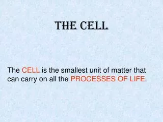
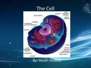
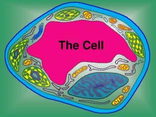
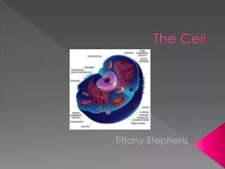
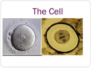
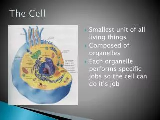
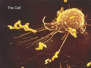
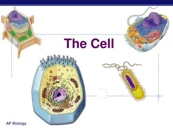
The Cell The cell
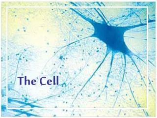
The cell is the functional unit of life What is the cell? On the other hand, there are diverse forms of life existing as single-celled organisms.
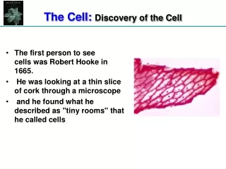
Microscopy • Although cells were discovered by Robert Hooke in 1665, the geography of the cell was largely uncharted until the past few decades.
Microscopy • The light microscope can resolve individual cells, it cannot however resolve much of the internal anatomy, especially the organelles. • To resolve smaller structures we use an electron microscope (EM), which focuses a beam of electrons through the specimen or onto its surface. • Theoretically, the resolution of a modern EM could reach 0.1 nanometer (nm), but the practical limit is closer to about 0.2 nm.
Transmission electron microscope (TEM) Transmission electron micrograph of cilia
Scanning electron microscope (SEM) Scanning electron micrograph of cilia
Important notes • Electron microscopes reveal organelles, but they can only be used on dead cells. • Light microscopes do not have as high a resolution, but they can be used to study live cells. • Microscopes are a major tool in cytology, the study of cell structures. • Cytology coupled with biochemistry (cell fractionation), the study of molecules and chemical processes in metabolism, developed into modern cell biology.
Cell fractionation • The goal of cell fractionation is to separate the major organelles of the cells so that their individual functions can be studied.
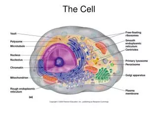
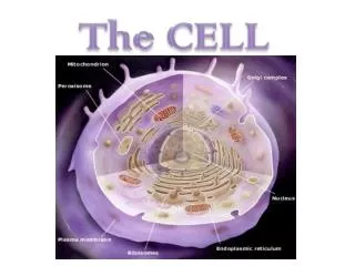
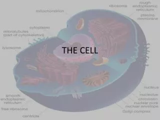
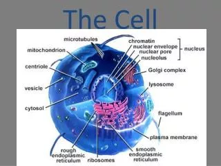
Structure and Function of the Cell • The cell is the basic functional unit of all living things. • The plasma membrane (cell membrane) bounds the cell and encloses the nucleusand cytoplasm. • The cytoplasmconsists of specialized bodies called organellessuspended in a fluid matrix, the cytosol, which consists of water and dissolved substances such as proteins and nutrients.
The plasma membrane • The plasma membraneseparates internal metabolic events from the external environment and controls the movement of materials into and out of the cell. • The plasma membrane is a double phospholipid membrane (lipid bilayer) with the polar hydrophilic heads forming the two outer faces and the nonpolar hydrophobic tails pointing toward the inside of the membrane
Proteins are scattered throughout the flexible phospholipid membrane. • Proteins may attach loosely to the inner or outer surface of the membrane (peripheral proteins), or they may extend into the membrane (integral proteins). Integral proteins may span across the membrane, appearing at both surfaces (transmembrane proteins).
Like phospholipids, integral proteins are amphipathic, with the hydrophobic regions embedded in the membrane and the hydrophilic regions exposed to the aqueous solutions bordering the membrane.
The mosaic nature of scattered proteins within a flexible matrix of phospholipid molecules describes the fluid mosaic modelof the cell membrane.
Additional features of the plasma membrane follow: • 1. The phospholipid membrane is selectively permeable. Only small, uncharged, polar molecules (such as H2O and CO2) and hydrophobic molecules (nonpolar molecules like O2 and lipid-soluble molecules such as hydrocarbons) freely pass across the membrane. • In contrast, large polar molecules (such as glucose) and all ions are impermeable.
2. Proteins in the plasma membrane provide a wide range of functions and include the following: • • Channel proteinsprovide passageways through the membrane for certain hydrophilic (water-soluble) substances such as polar and charged molecules. • • Transport proteinsspend energy (ATP) to transfer materials across the membrane. When energy is used for this purpose, the materials are said to be activelytransported, and the process is called active transport.
• Recognition proteinsdistinguish the identity of neighboring cells. These proteins are glycoproteinsbecause they have short polysaccharide chains (oligosaccharides) attached. The oligosaccharide part of the glycoprotein protrudes from the surface of the membrane like an antenna. • • Adhesion proteinsattach cells to neighboring cells or provide anchors for the internal filaments and tubules that give stability to the cell. oligosaccharides Adhesion proteins Recognition proteins
Receptor proteinsprovide binding sites for hormones or other trigger molecules. In response to the hormone or trigger molecule, a specific cell response is activated. • Electron transfer proteinsare involved in transferring electrons from one molecule to another during chemical reactions.
Cholesterol Cholesterol Cholesterol Cholesterol Cholesterol Cholesterol 3. Cholesterolmolecules distributed throughout the phospholipid bilayer provide some rigidity to the plasma membranes of animal cells. In plant cells, related substances (sterols) provide a similar function. CYTOPLASM Cholesterol helps maintain the fluidity of the membrane by preventing the phospholipids from packing too tightly.
The glycocalyx • 4. The glycocalyxis a carbohydrate “coat” covering the outer face of the plasma membrane. It consists of various oligosaccharides that are attached to membrane phospholipids (glycolipids) and proteins (such as the glycoproteinsof recognition proteins). The glycocalyx provides markers for cell-cell recognition.
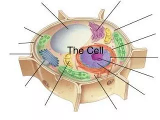
Organelles • Organelles are bodies within the cytoplasm that serve to physically separate the various metabolic reactions that occur within cells.
NUCLEUS Chromatin Two membranesof nuclearenvelope Nucleolus Pore ROUGHENDOPLASMICRETICULUM Ribosomes 1. The nucleus • The nucleus is bounded by the nuclear envelope, a phospholipid bilayer similar to the plasma membrane. • The nucleus contains DNA (deoxyribonucleic acid), the hereditary information of the cell. Normally, the DNA is spread out within the nucleus as a threadlike matrix called chromatin.
DNAdoublehelix(2-nmdiameter) 1. The nucleus Histones “Beads ona string” • When the cell begins to divide, the chromatin condenses into rod-shaped bodies called chromosomes, each of which, before dividing, is made up of two long DNA molecules and various histone (protein) molecules. The histones serve to organize the lengthy DNA, coiling it into bundles called nucleosomes. Nucleosome(10-nm diameter) Tight helical fiber(30-nm diameter) Supercoil(100-300nm diameter) 700nm Metaphase chromosome
NUCLEUS Chromatin Two membranesof nuclearenvelope Nucleolus Pore ROUGHENDOPLASMICRETICULUM Ribosomes 1. The nucleus • Also visible within the nucleus are one or more nucleoli, concentrations of DNA in the process of manufacturing the components of ribosomes. • The nucleus also serves as the site for the separation of chromosomes during cell division.
2. Ribosome • Ribosome subunits are manufactured in the nucleus and consist of RNA molecules and proteins. • The two subunits, labeled 60S and 40S, move across the nuclear envelope and into the cytoplasm where they are assembled into a single 80S ribosome. • (An S value, or Svedberg unit, expresses how readily a product forms a sediment in a centrifuge, with larger values representing larger and heavier products).
In the cytoplasm, ribosomes assist in the assembly of amino acids into proteins.
SMOOTH ER ROUGHER Nuclearenvelope Ribosomes SMOOTH ER ROUGH ER 3. The endoplasmic reticulum (ER) • The endoplasmic reticulum, or ER, consists of stacks of flattened sacs involved in the production of various materials. • In cross section, they appear as a series of maze-like channels, often closely associated with the nucleus.
When ribosomes are present, the ER (called rough ER) creates glycoproteinsby attaching polysaccharide groups to polypeptides as they are assembled by the ribosomes.
Smooth ER, without ribosomes, is responsible for various activities, including the synthesis of lipids and hormones, especially in cells that produce these substances for export from the cell. • In liver cells, smooth ER is involved in the breakdown of toxins, drugs, and toxic by-products from cellular reactions.
4. A Golgi apparatus (Golgi complex or Golgi body) • A Golgi apparatus (Golgi complex or Golgi body) is a group of flattened sacs arranged like a stack of bowls. • They function to modify and package proteins and lipids into vesicles, small, spherically shaped sacs that bud from the outside surface of the Golgi apparatus.
Transport vesiclefrom Golgi Transport vesiclefrom ER Rough ER Plasmamembrane Vacuole Nucleus Lysosome Golgiapparatus Smooth ER Nuclearenvelope • Vesicles often migrate to and merge with the plasma membrane, releasing their contents to the outside of the cell.
Vesicles often migrate to and merge with the plasma membrane, releasing their contents to the outside of the cell.
5. Lysosomes • Lysosomes are vesicles from a Golgi apparatus that contain digestive enzymes.
Rough ER Transport vesicle(containing inactivehydrolytic enzymes) Plasmamembrane Golgiapparatus Engulfmentof particle Lysosomeengulfingdamagedorganelle “Food” LYSOSOMES Digestion Foodvacuole • They break down food, cellular debris, and foreign invaders such as bacteria. • Lysosomes do not occur in plant cells.
6. Peroxisomes • Peroxisomes are organelles that break down various substances. During the breakdown process, O2 combines with hydrogen to form toxic hydrogen peroxide (H2O2), which in turn is converted to H2O. • Peroxisomes are common in liver and kidney cells where they break down toxic substances and in photosynthesizing plant cells. Peroxisomes transfer hydrogen from various substances to oxygen
7. Mitochondria • Mitochondria carry out aerobic respiration, a process in which energy (in the form of ATP) is obtained from carbohydrates.
8. Chloroplasts • Chloroplasts carry out photosynthesis, the plant process of incorporating energy from sunlight into carbohydrates.
Tubulinsubunit Actin subunit Fibrous subunits 25 nm 7 nm 10 nm INTERMEDIATEFILAMENT MICROFILAMENT MICROTUBULE 9. Microtubules, intermediate filaments, and microfilaments • Microtubules, intermediate filaments, and microfilaments are three protein fibers of decreasing diameter, respectively. • All are involved in establishing the shape of or in coordinating movements of the cytoskeleton, the internal structure of the cytoplasm.
Microtubules • Microtubules are made of the protein tubulin and provide support and motility for cellular activities. • They are found in the spindle apparatus (which guides the movement of chromosomes during cell division), and in flagella and cilia, structures that project from the plasma membrane to provide motility to the cell.
Intermediate filaments • Intermediate filaments provide support for maintaining the shape of the cell.
phagocytes Microfilaments • Microfilaments are made of the protein actinand are involved in cell motility. • They are found in muscle cells and in cells that move by changing shape, such as phagocytes (white blood cells that wander throughout the body attacking bacteria and other foreign invaders).
10. Flagella and cilia • Flagella and cilia are structures that protrude from the cell membrane and make wavelike movements. • Flagella and cilia are classified by their lengths and by their numbers per cell: • flagella are long and few; cilia are short and many. A single flagellum propels sperm, while the numerous cilia that line the respiratory tract sweep away debris.
10. Flagella and cilia • Structurally, both flagella and cilia consist of microtubules arranged in a “9 + 2” array–nine pairs (doublets) of microtubules arranged in a circle surrounding a pair of microtubules.
FLAGELLUM Electron micrograph of sections: Outer microtubule doublet Plasmamembrane Flagellum Centralmicrotubules Outer microtubule doublet Plasmamembrane Basal body Basal body(structurally identical to centriole)
INTERPHASE PROPHASE Centrosomes(with centriole pairs) Early mitoticspindle Centrosome Fragmentsof nuclearenvelope Kinetochore Chromatin Centrosome Spindlemicrotubules Nucleolus Nuclearenvelope Plasmamembrane Chromosome,consisting of twosister chromatids 11. Centrioles and basal bodies • Centrioles and basal bodies act as microtubule organizing centers (MTOCs). • A pair of centrioles (enclosed in a centrosome) located outside the nuclear envelope gives rise to the microtubules that make up the spindle apparatus used during cell division.
Basal bodies are at the base of each flagellum and cilium and appear to organize their development. • Both centrioles and basal bodies are made up of nine triplets arranged in a circle.
Life cycle of Ferns السَّرْخَس Life cycle of Mosses حَزازُ • Plant cells lack centrioles and only “lower” plants (such as mosses and ferns) with motile sperm have flagella and basal bodies.