RED EYE
RED EYE. Maria Isabel Diaz, MD St. Barnabas Hospital Department of Pediatrics 1/7/2010. Objectives. Develop a DDx for Red Eye Be able to differentiate between serious, vision-threatening conditions and benign conditions that cause a Red Eye. Anatomy of the Eye. Anatomy of the Eye.
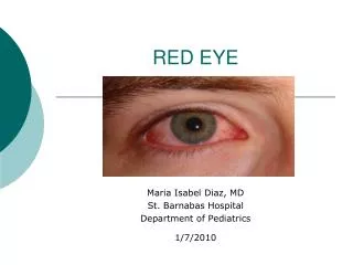

RED EYE
E N D
Presentation Transcript
RED EYE Maria Isabel Diaz, MD St. Barnabas Hospital Department of Pediatrics 1/7/2010
Objectives • Develop a DDx for Red Eye • Be able to differentiate between serious, vision-threatening conditions and benign conditions that cause a Red Eye.
Red Eye • Cardinal sign of ocular inflammation. • Most cases benign and can be managed by PCP. • Key to management is recognizing cases with underlying disease that require consultation.
Pathophysiology • The red eye is caused by the dilation of blood vessels in the eye. • Should differentiate between ciliary and conjunctival injection.
Pathophysiology • Ciliary injection: involves branches of the anterior ciliary arteries. • Indicates inflammation of the cornea, iris or ciliary body.
Pathophysiology • Conjunctival Injection: mainly affects the posterior conjunctival blood vessels. • Because these vessels are more superficial than the ciliary arteries, they produce more redness and constrict with vasoconstrictors.
Prior episodes Ophthalmologic history including eye sx Bilateral or unilateral Contact lens use Comorbid conditions Onset Visual changes Trauma Photophobia Pain Discharge, clear or colored Clinical: History
Visual acuity Extraocular movements Pupil reactivity Pupil shape Photophobia Slit lamp examination with and without fluorescein * IOP measurements * Eyelid inspection with eversion Clinical: Physical
No Pain and normal vision Likely to have self-limiting condition. Conjunctivitis Episcleritis Subconjunctival hemorrhage Pain with/out blurring of vision Likely to have a sight-threatening condition: Acute glaucoma Iritis Corneal infections Causes of Red Eye
Conjunctivitis • Characterized by vascular dilation, cellular infiltration and exudation. Allergic: Often papillary projections and pruritus. + h/o allergic ds. Viral: + lymphoid follicles on the undersurface of the lid and enlarged tender pre-auricular nodes.
Conjunctivitis Bacterial: More purulent disease. Differentiating the three types is not easy, when unclear assume that a bacterial etiology is involved.
Conjunctivitis Follicles Purulent discharge Papillae Chemosis Redness
Conjunctivitis • Treatment: • In the general practice, it is difficult to differentiate between bacterial from viral conjunctivitis. It is acceptable to treat all infective conjunctivitis with topical antibiotics as it can prevent secondary infection in viral conjunctivitis. • Patient with allergic conjunctivitis will benefit from topical allergy drops. • Oral antihistamine is useful in reducing itchiness. It is important to determine the cause. • Refer the patient to the specialist only if the conjunctivitis fails to respond to treatment
Episcleritis • Superficial • Idiopathic, but R/o collagen vascular disorder. • Asymptomatic, mild pain • Self-limiting or topical treatment • H/o recurrent episodes is common
Episcleritis • Management: • This condition is self-limiting • If there is no discomfort, no treatment is needed. • The condition resolves within two weeks. • If the patient complains of discomfort or if the problem fails to resolve spontaneously, refer the patient in the same week. Topical mild steroid may be needed.
Subconjunctival Haemorrhage • Diffuse or localized area of blood under conjunctiva. • Asymptomatic • Idiopathic, trauma, cough, sneezing, aspirin, HT • Resolves within 10-14 days
Subconjunctival Haemorrhage • Management: • The condition looks alarming but resolves within two weeks. • Reassurance is all that is needed. • Refer the patient only if the subjconjunctival hemorrhage is traumatic.
Foreign Body • Eye should be stained with fluorescein to detect evidence of corneal abrasion. • Penetration of the globe should be excluded by thorough slit lamp examination. • The lid should always be everted to exclude retained material.
Blepharitis • Inflammation of the eyelids usually involving the lid margins. • Often associated with conjunctivitis • May be seborrheic or caused by staphylococcal infection.
Canaliculitis • Mildly red eye (usually unilateral) • Slight discharge, can be expressed from the canaliculus. • Often is caused by Actinomyces israelli.
Corneal Inflammation or Infection • May have decrease visual acuityand photophobia. • Often c/o severe pain • Epithelial defect may be evident on slit lamp examination or may require staining with fluorescein. • ANY opacification of the cornea in a red eye is an infection of the cornea until proven otherwise. • THIS IS AN OPHTHALMOLOGIC EMERGENCY.
Corneal Infections • Management: • Refers within 24 hours • In herpes keratitis, topical acyclovir 3% five times a day is prescribed for one week • In bacterial corneal ulcer, the patient may be admitted for intensive antibiotic treatment if severe or treated as an out-patient if mild
Corneal Abrasion • Surface epithelium sloughed off. • Stains with fluorescein • Usually due to trauma • Pain, FB sensation,tearing, red eye.
Corneal Ulcer • Infection • Bacterial • Viral • Fungal • Protozoan • Mechanical or trauma • Chemical: Alkali injuries are worse than acid
The picture shows a corneal ulcer with hypopyon. Refer urgently.
Fluorescein staining reveals a dendritic ulcer typical of Herpes keratitis. This is treated with topical 3% acyclovir.
Scleritis • Deep • Idiopathic • Painful, gradual onset of red eye, insidious decrease in vision. • Globe is often tender and sclera swollen. • A deep violet discoloration may be observed (dilation of deep venous plexus) • Collagen vascular disease, Zoster, Sarcoidosis • Systemic treatment with NSAI or Prednisolone if severe
Anterior uveitis (iritis) • Photophobia, perilimbal injection, decreased vision • Idiopathic- most common. • Associated to systemic disease • Seronegative arthropathies:AS, IBD, Psoriatic arthritis, Reiter’s • Autoimmune: Sarcoidosis, Behcets • Infection: Shingles, Toxoplasmosis, TB, Syphillis, HIV
Painful photophobic Red eye. Note the ciliary injection around the cornea (limbus) typical of iritis
Iritis • Management: • Refer the patient within 24 hours. • Slit-lamp examination by ophthalmologists to confirm the diagnosis. • Treatment is with intensive topical steroid to reduce inflammation and mydriatic to dilate the pupil so that the iris does not stick to the cornea causing problem with glaucoma.
Acute Angle-closure Glaucoma • Symptoms • Pain, headache, nausea-vomiting • Redness, photophobia, • Reduced vision • Haloes around lights • Patient usually older than 50 y • IOP increased Ciliary hyperemia Dilated pupil Corneal edema
Acute Angle-closure Glaucoma • Management: • Urgent referrals as soon as possible and not the next day. • Patient is usually admitted and given mannitol IV to lower pressure. Topical pilocarpine and steroid (to reduce inflammation) are also given.
Summary • Red eye is a common complaint. • Bad signs - REFER • Decrease VA • Abnormalities with Fluorescein staining. • Unequal size or unreactive pupil. • Proptosis • Ciliary flush • Corneal opacities • Limited or painful EOM • Increase IOP • Cases requiring prolonged treatment or who do not respond as expected to the treatment.